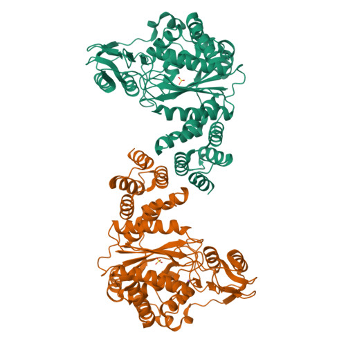
X-Ray Structure of the Complex Pyrimidine-nucleoside phosphorylase from Bacillus subtilis with Sulfate Ion
Lashkov, A.A., Balaev, V.V., Gabdoulkhakov, A.G., Betzel, C., Mikhailov, A.M.To be published.
Experimental Data Snapshot
Starting Model: experimental
View more details
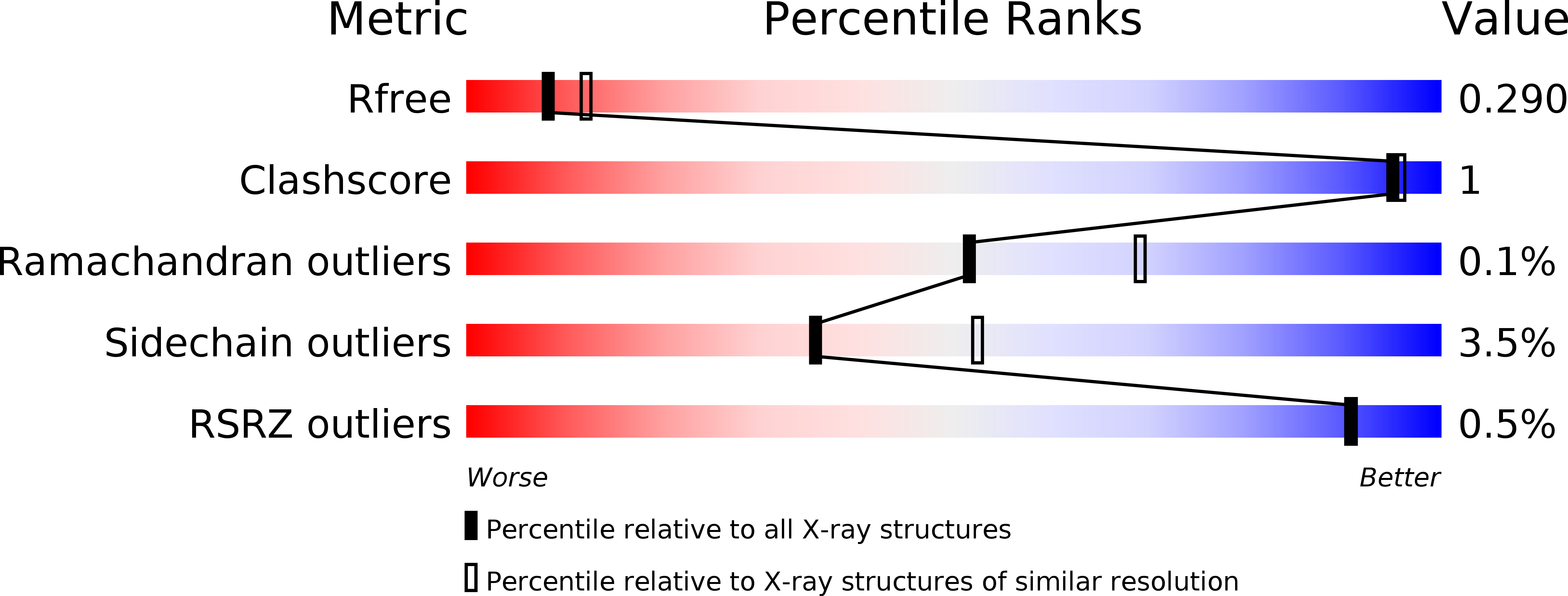
wwPDB Validation 3D Report Full Report
Entity ID: 1 | |||||
|---|---|---|---|---|---|
| Molecule | Chains | Sequence Length | Organism | Details | Image |
| Pyrimidine-nucleoside phosphorylase | 433 | Bacillus subtilis subsp. subtilis str. 168 | Mutation(s): 0 Gene Names: pdp, BSU39400 EC: 2.4.2.2 | 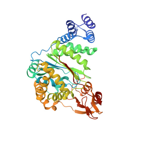 | |
UniProt | |||||
Find proteins for P39142 (Bacillus subtilis (strain 168)) Explore P39142 Go to UniProtKB: P39142 | |||||
Entity Groups | |||||
| Sequence Clusters | 30% Identity50% Identity70% Identity90% Identity95% Identity100% Identity | ||||
| UniProt Group | P39142 | ||||
Sequence AnnotationsExpand | |||||
| |||||
| Ligands 2 Unique | |||||
|---|---|---|---|---|---|
| ID | Chains | Name / Formula / InChI Key | 2D Diagram | 3D Interactions | |
| SO4 Query on SO4 | C [auth A], D [auth B] | SULFATE ION O4 S QAOWNCQODCNURD-UHFFFAOYSA-L |  | ||
| NA Query on NA | E [auth B] | SODIUM ION Na FKNQFGJONOIPTF-UHFFFAOYSA-N |  | ||
| Length ( Å ) | Angle ( ˚ ) |
|---|---|
| a = 80.463 | α = 90 |
| b = 91.35 | β = 90 |
| c = 109.8 | γ = 90 |
| Software Name | Purpose |
|---|---|
| REFMAC | refinement |
| SCALA | data scaling |
| MOLREP | phasing |
| PDB_EXTRACT | data extraction |
| XDS | data reduction |
| Funding Organization | Location | Grant Number |
|---|---|---|
| RFBR | Russian Federation | 14-04-00952 |