5EIN

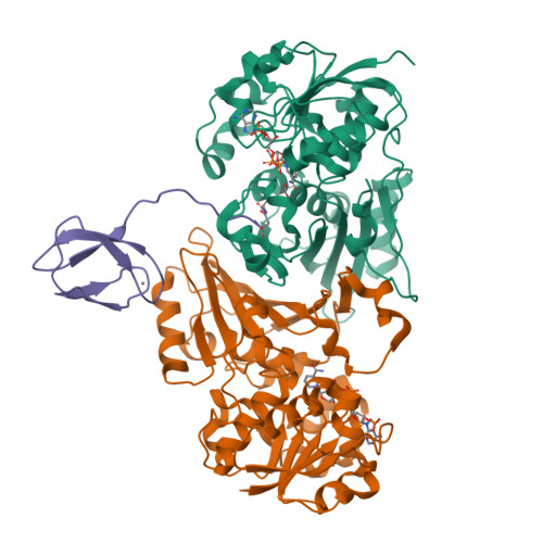
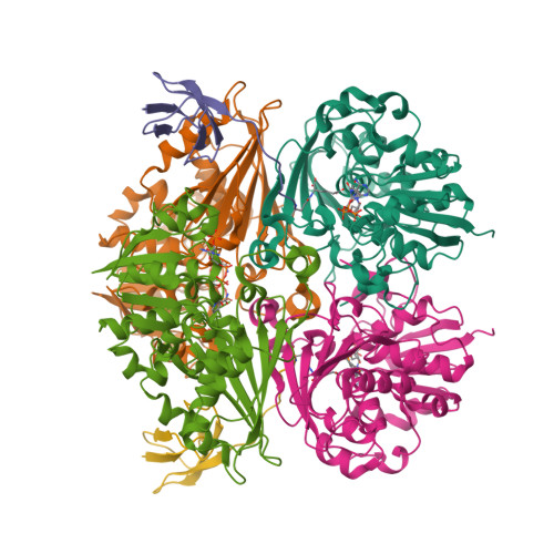
Crystal structure of C148A mutant of LysY from Thermus thermophilus in complex with NADP+ and LysW-gamma-aminoadipic acid


Experimental Data Snapshot

  • Method: X-RAY DIFFRACTION
  • Resolution: 1.70 Å
  • R-Value Free: 0.210 
  • R-Value Work: 0.171 
  • R-Value Observed: 0.173 

Starting Model: experimental
View more details

wwPDB Validation   3D Report Full Report


Ligand Structure Quality Assessment 


This is version 3.1 of the entry. See complete history


Literature

Crystal Structure of the LysYLysW Complex from Thermus thermophilus.

Shimizu, T.Tomita, T.Kuzuyama, T.Nishiyama, M.

(2016) J Biol Chem 291: 9948-9959

  • DOI: https://doi.org/10.1074/jbc.M115.707034
  • Primary Citation of Related Structures:  
    5EIN, 5EIO

  • PubMed Abstract: 

    Several bacteria and archaea utilize the amino group-carrier protein, LysW, for lysine biosynthesis, in which an isopeptide bond is formed between the C-terminal Glu of LysW and an amino group of α-aminoadipate (AAA). The resulting LysW-γ-AAA is phosphorylated by LysZ to form LysW-γ-AAA phosphate, which is subsequently reduced to LysW-γ-aminoadipic semialdehyde (LysW-γ-AASA) through a reaction catalyzed by LysY. In this study, we determined the crystal structures of LysY from Thermus thermophilus HB27 (TtLysY) complexed with TtLysW-γ-AASA and TtLysW-γ-AAA, respectively. In both structures, the globular domain of TtLysW was recognized by positively charged residues on helix α9 and the β11-α10 loop of TtLysY through conformational changes. A mutational analysis confirmed that the interactions observed between TtLysY and TtLysW are important for the function of TtLysY. The extended LysW recognition loop and conserved arginine residue were identified as signatures to discriminate LysY from ArgC, which is involved in arginine biosynthesis. Combined with the previously determined TtLysZ·TtLysW complex structure, TtLysW may simultaneously bind TtLysZ and TtLysY. These structural insights suggest the formation of a TtLysWZY ternary complex, in which the flexible C-terminal extension of TtLysW promotes the efficient transfer of the labile intermediate from the active site of TtLysZ to that of TtLysY during the sequential reactions catalyzed by TtLysZY.


  • Organizational Affiliation

    From the Biotechnology Research Center, University of Tokyo, 1-1-1, Yayoi, Bunkyo-ku, Tokyo 113-8657, Japan.


Macromolecules
Find similar proteins by:  (by identity cutoff)  |  3D Structure
Entity ID: 1
MoleculeChains Sequence LengthOrganismDetailsImage
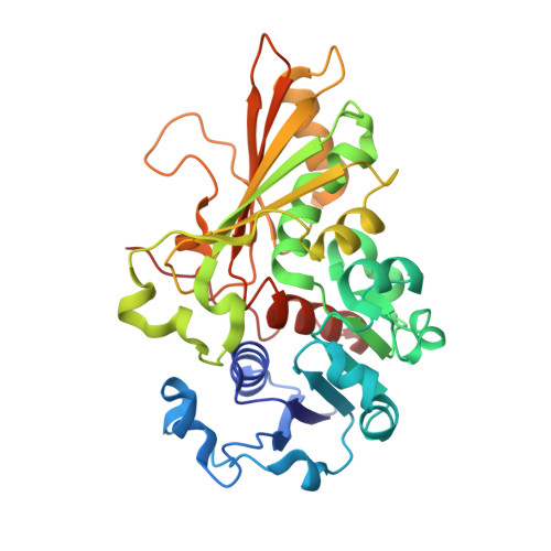
N-acetyl-gamma-glutamyl-phosphate/N-acetyl-gamma-aminoadipyl-phosphate reductase
A, B
344Thermus thermophilus HB27Mutation(s): 1 
Gene Names: argC2argClysYTT_C1542
EC: 1.2.1 (PDB Primary Data), 1.2.1.38 (PDB Primary Data), 1.2.1.103 (UniProt)
UniProt
Find proteins for O50146 (Thermus thermophilus (strain ATCC BAA-163 / DSM 7039 / HB27))
Explore O50146 
Go to UniProtKB:  O50146
Entity Groups  
Sequence Clusters30% Identity50% Identity70% Identity90% Identity95% Identity100% Identity
UniProt GroupO50146
Sequence Annotations
Expand
  • Reference Sequence
Find similar proteins by:  (by identity cutoff)  |  3D Structure
Entity ID: 2
MoleculeChains Sequence LengthOrganismDetailsImage
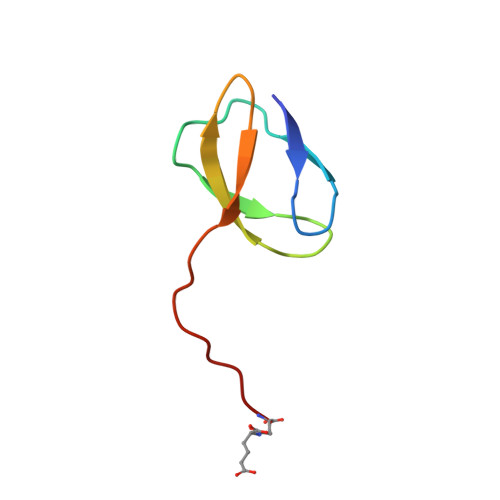
Alpha-aminoadipate carrier protein LysW54Thermus thermophilusMutation(s): 0 
Gene Names: orfF
UniProt
Find proteins for Q9ZND7 (Thermus thermophilus)
Explore Q9ZND7 
Go to UniProtKB:  Q9ZND7
Entity Groups  
Sequence Clusters30% Identity50% Identity70% Identity90% Identity95% Identity100% Identity
UniProt GroupQ9ZND7
Sequence Annotations
Expand
  • Reference Sequence
Small Molecules
Ligands 3 Unique
IDChains Name / Formula / InChI Key2D Diagram3D Interactions
NAP
Query on NAP

Download Ideal Coordinates CCD File 
D [auth A],
Q [auth B]
NADP NICOTINAMIDE-ADENINE-DINUCLEOTIDE PHOSPHATE
C21 H28 N7 O17 P3
XJLXINKUBYWONI-NNYOXOHSSA-N
ZN
Query on ZN

Download Ideal Coordinates CCD File 
CA [auth C]ZINC ION
Zn
PTFCDOFLOPIGGS-UHFFFAOYSA-N
FMT
Query on FMT

Download Ideal Coordinates CCD File 
AA [auth B]
BA [auth B]
E [auth A]
F [auth A]
G [auth A]
AA [auth B],
BA [auth B],
E [auth A],
F [auth A],
G [auth A],
H [auth A],
I [auth A],
J [auth A],
K [auth A],
L [auth A],
M [auth A],
N [auth A],
O [auth A],
P [auth A],
R [auth B],
S [auth B],
T [auth B],
U [auth B],
V [auth B],
W [auth B],
X [auth B],
Y [auth B],
Z [auth B]
FORMIC ACID
C H2 O2
BDAGIHXWWSANSR-UHFFFAOYSA-N
Modified Residues  1 Unique
IDChains TypeFormula2D DiagramParent
R0K
Query on R0K
C
L-PEPTIDE LINKINGC11 H18 N2 O7GLU
Experimental Data & Validation

Experimental Data

  • Method: X-RAY DIFFRACTION
  • Resolution: 1.70 Å
  • R-Value Free: 0.210 
  • R-Value Work: 0.171 
  • R-Value Observed: 0.173 
  • Space Group: P 32 2 1
Unit Cell:
Length ( Å )Angle ( ˚ )
a = 83.54α = 90
b = 83.54β = 90
c = 167.781γ = 120
Software Package:
Software NamePurpose
REFMACrefinement
Cootmodel building
HKL-2000data scaling
HKL-2000data collection
PHASERphasing

Structure Validation

View Full Validation Report


Ligand Structure Quality Assessment 


Entry History 

Deposition Data

Revision History  (Full details and data files)

  • Version 1.0: 2016-03-23
    Type: Initial release
  • Version 1.1: 2020-02-19
    Changes: Data collection, Database references, Derived calculations
  • Version 2.0: 2023-04-05
    Changes: Atomic model, Data collection, Database references, Derived calculations, Non-polymer description, Other, Polymer sequence, Refinement description, Structure summary
  • Version 3.0: 2023-11-15
    Changes: Atomic model, Data collection, Derived calculations
  • Version 3.1: 2023-11-29
    Changes: Refinement description