5EGF

The crystal structure of SeMet-CT


Experimental Data Snapshot

  • Method: X-RAY DIFFRACTION
  • Resolution: 2.29 Å
  • R-Value Free: 0.266 
  • R-Value Work: 0.225 
  • R-Value Observed: 0.226 

wwPDB Validation   3D Report Full Report


Ligand Structure Quality Assessment 


This is version 1.2 of the entry. See complete history


Literature

Structural basis of nonribosomal peptide macrocyclization in fungi

Zhang, J.Liu, N.Cacho, R.A.Gong, Z.Liu, Z.Qin, W.Tang, C.Tang, Y.Zhou, J.

(2016) Nat Chem Biol 12: 1001-1003

  • DOI: https://doi.org/10.1038/nchembio.2202
  • Primary Citation of Related Structures:  
    5DIJ, 5DLK, 5EGF, 5EJD

  • PubMed Abstract: 

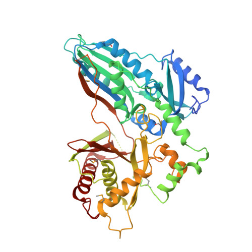
    Nonribosomal peptide synthetases (NRPSs) in fungi biosynthesize important pharmaceutical compounds, including penicillin, cyclosporine and echinocandin. To understand the fungal strategy of forging the macrocyclic peptide linkage, we determined the crystal structures of the terminal condensation-like (C T ) domain and the holo thiolation (T)-C T complex of Penicillium aethiopicum TqaA. The first, to our knowledge, structural depiction of the terminal module in a fungal NRPS provides a molecular blueprint for generating new macrocyclic peptide natural products.


  • Organizational Affiliation

    State Key Laboratory of Bio-organic and Natural Products Chemistry, Shanghai Institute of Organic Chemistry, Chinese Academy of Sciences, Shanghai 200032, China.


Macromolecules
Find similar proteins by:  (by identity cutoff)  |  3D Structure
Entity ID: 1
MoleculeChains Sequence LengthOrganismDetailsImage
TqaA
A, B, C, D
486Penicillium aethiopicumMutation(s): 2 
UniProt
Find proteins for F1CWE4 (Penicillium aethiopicum)
Explore F1CWE4 
Go to UniProtKB:  F1CWE4
Entity Groups  
Sequence Clusters30% Identity50% Identity70% Identity90% Identity95% Identity100% Identity
UniProt GroupF1CWE4
Sequence Annotations
Expand
  • Reference Sequence
Small Molecules
Ligands 2 Unique
IDChains Name / Formula / InChI Key2D Diagram3D Interactions
C8E
Query on C8E

Download Ideal Coordinates CCD File 
FA [auth C],
L [auth A],
QA [auth D],
W [auth B]
(HYDROXYETHYLOXY)TRI(ETHYLOXY)OCTANE
C16 H34 O5
FEOZZFHAVXYAMB-UHFFFAOYSA-N
EDO
Query on EDO

Download Ideal Coordinates CCD File 
AA [auth C]
BA [auth C]
CA [auth C]
DA [auth C]
E [auth A]
AA [auth C],
BA [auth C],
CA [auth C],
DA [auth C],
E [auth A],
EA [auth C],
F [auth A],
G [auth A],
GA [auth D],
H [auth A],
HA [auth D],
I [auth A],
IA [auth D],
J [auth A],
JA [auth D],
K [auth A],
KA [auth D],
LA [auth D],
M [auth B],
MA [auth D],
N [auth B],
NA [auth D],
O [auth B],
OA [auth D],
P [auth B],
PA [auth D],
Q [auth B],
R [auth B],
S [auth B],
T [auth B],
U [auth B],
V [auth B],
X [auth C],
Y [auth C],
Z [auth C]
1,2-ETHANEDIOL
C2 H6 O2
LYCAIKOWRPUZTN-UHFFFAOYSA-N
Modified Residues  1 Unique
IDChains TypeFormula2D DiagramParent
MSE
Query on MSE
A, B, C, D
L-PEPTIDE LINKINGC5 H11 N O2 SeMET
Experimental Data & Validation

Experimental Data

  • Method: X-RAY DIFFRACTION
  • Resolution: 2.29 Å
  • R-Value Free: 0.266 
  • R-Value Work: 0.225 
  • R-Value Observed: 0.226 
  • Space Group: C 1 2 1
Unit Cell:
Length ( Å )Angle ( ˚ )
a = 216.602α = 90
b = 100.284β = 100.18
c = 128.23γ = 90
Software Package:
Software NamePurpose
PHENIXrefinement
PHENIXphasing
AutoSolmodel building
HKL-2000data processing

Structure Validation

View Full Validation Report


Ligand Structure Quality Assessment 


Entry History 

Deposition Data

Revision History  (Full details and data files)

  • Version 1.0: 2016-10-19
    Type: Initial release
  • Version 1.1: 2016-11-30
    Changes: Database references
  • Version 1.2: 2020-02-19
    Changes: Data collection