4YH2 | pdb_00004yh2

Glutathione Transferase E6 from Drosophila melanogaster


Experimental Data Snapshot

  • Method: X-RAY DIFFRACTION
  • Resolution: 1.72 Å
  • R-Value Free: 
    0.188 (Depositor), 0.190 (DCC) 
  • R-Value Work: 
    0.157 (Depositor), 0.160 (DCC) 
  • R-Value Observed: 
    0.159 (Depositor) 

wwPDB Validation   3D Report Full Report


Ligand Structure Quality Assessment 

Created with Raphaël 2.3.0Worse 01 BetterLigand structure goodness of fit to experimental dataBest fitted GSHClick on this verticalbar to view details

This is version 1.2 of the entry. See complete history


Literature

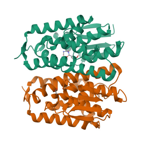
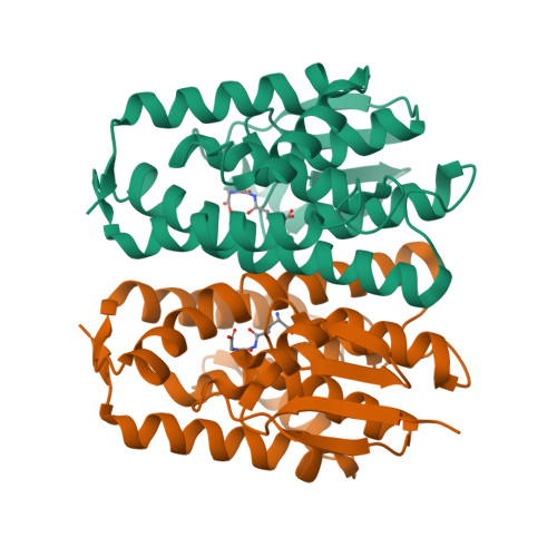
Epsilon glutathione transferases possess a unique class-conserved subunit interface motif that directly interacts with glutathione in the active site

Wongsantichon, J.Robinson, R.C.Ketterman, A.J.

(2015) Biosci Rep 35

  • DOI: https://doi.org/10.1042/BSR20150183
  • Primary Citation of Related Structures:  
    4YH2

  • PubMed Abstract: 

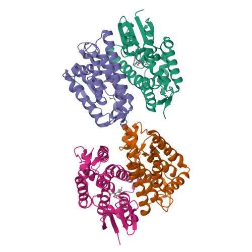
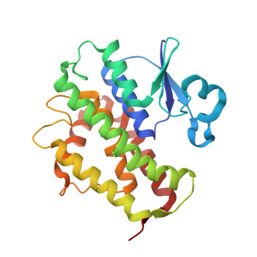
    Epsilon class glutathione transferases (GSTs) have been shown to contribute significantly to insecticide resistance. We report a new Epsilon class protein crystal structure from Drosophila melanogaster for the glutathione transferase DmGSTE6. The structure reveals a novel Epsilon clasp motif that is conserved across hundreds of millions of years of evolution of the insect Diptera order. This histidine-serine motif lies in the subunit interface and appears to contribute to quaternary stability as well as directly connecting the two glutathiones in the active sites of this dimeric enzyme.


  • Organizational Affiliation

    Institute of Molecular and Cell Biology, A*STAR (Agency for Science, Technology and Research), Biopolis, Singapore 138673.


Macromolecules
Find similar proteins by:  (by identity cutoff)  |  3D Structure
Entity ID: 1
MoleculeChains Sequence LengthOrganismDetailsImage
Glutathione S transferase E6
A, B, C, D
222Drosophila melanogasterMutation(s): 0 
Gene Names: GstE6CG17530Dmel_CG17530
EC: 2.5.1.18
UniProt
Find proteins for A1ZB71 (Drosophila melanogaster)
Explore A1ZB71 
Go to UniProtKB:  A1ZB71
Entity Groups  
Sequence Clusters30% Identity50% Identity70% Identity90% Identity95% Identity100% Identity
UniProt GroupA1ZB71
Sequence Annotations
Expand
  • Reference Sequence
Experimental Data & Validation

Experimental Data

  • Method: X-RAY DIFFRACTION
  • Resolution: 1.72 Å
  • R-Value Free:  0.188 (Depositor), 0.190 (DCC) 
  • R-Value Work:  0.157 (Depositor), 0.160 (DCC) 
  • R-Value Observed: 0.159 (Depositor) 
Space Group: C 1 2 1
Unit Cell:
Length ( Å )Angle ( ˚ )
a = 176.35α = 90
b = 58.86β = 128.25
c = 122.81γ = 90
Software Package:
Software NamePurpose
Aimlessdata scaling
PHENIXrefinement
PDB_EXTRACTdata extraction
xia2data reduction
xia2phasing

Structure Validation

View Full Validation Report


Ligand Structure Quality Assessment 

Created with Raphaël 2.3.0Worse 01 BetterLigand structure goodness of fit to experimental dataBest fitted GSHClick on this verticalbar to view details

Entry History 

Deposition Data

Revision History  (Full details and data files)

  • Version 1.0: 2016-02-10
    Type: Initial release
  • Version 1.1: 2020-02-05
    Changes: Data collection, Database references, Derived calculations
  • Version 1.2: 2024-03-20
    Changes: Data collection, Database references