4R8Z

Crystal Structure of PA4781 HD-GYP domain from Pseudomonas aeruginosa at 2.2A resolution showing a bi-metallic Ni ion center


Experimental Data Snapshot

  • Method: X-RAY DIFFRACTION
  • Resolution: 2.20 Å
  • R-Value Free: 0.213 
  • R-Value Work: 0.177 
  • R-Value Observed: 0.179 

wwPDB Validation   3D Report Full Report


This is version 1.2 of the entry. See complete history


Literature

Structural basis of functional diversification of the HD-GYP domain revealed by the Pseudomonas aeruginosa PA4781 protein, which displays an unselective bimetallic binding site.

Rinaldo, S.Paiardini, A.Stelitano, V.Brunotti, P.Cervoni, L.Fernicola, S.Protano, C.Vitali, M.Cutruzzola, F.Giardina, G.

(2015) J Bacteriol 197: 1525-1535

  • DOI: https://doi.org/10.1128/JB.02606-14
  • Primary Citation of Related Structures:  
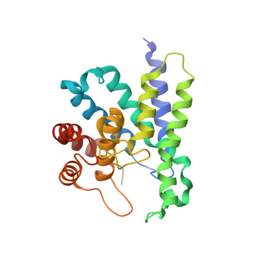
    4R8Z

  • PubMed Abstract: 

    The intracellular level of the bacterial secondary messenger cyclic di-3',5'-GMP (c-di-GMP) is determined by a balance between its biosynthesis and degradation, the latter achieved via dedicated phosphodiesterases (PDEs) bearing a characteristic EAL or HD-GYP domain. We here report the crystal structure of PA4781, one of the three Pseudomonas aeruginosa HD-GYP proteins, which we have previously characterized in vitro. The structure shows a bimetallic active site whose metal binding mode is different from those of both HD-GYP PDEs characterized so far. Purified PA4781 does not contain iron in the active site as for other HD-GYPs, and we show that it binds to a wide range of transition metals with similar affinities. Moreover, the structural features of PA4781 indicate that this is preferentially a pGpG binding protein, as we previously suggested. Our results point out that the structural features of HD-GYPs are more complex than predicted so far and identify the HD-GYP domain as a conserved scaffold which has evolved to preferentially interact with a partner GGDEF but which harbors different functions obtained through diversification of the active site.


  • Organizational Affiliation

    Istituto Pasteur-Fondazione Cenci Bolognetti, Department of Biochemical Sciences, Sapienza University of Rome, Rome, Italy.


Macromolecules
Find similar proteins by:  (by identity cutoff)  |  3D Structure
Entity ID: 1
MoleculeChains Sequence LengthOrganismDetailsImage
Cyclic di-GMP phosphodiesterase
A, B
218Pseudomonas aeruginosa PAO1Mutation(s): 0 
Gene Names: PA4781
EC: 3.1.4.1
UniProt
Find proteins for Q9HV27 (Pseudomonas aeruginosa (strain ATCC 15692 / DSM 22644 / CIP 104116 / JCM 14847 / LMG 12228 / 1C / PRS 101 / PAO1))
Explore Q9HV27 
Go to UniProtKB:  Q9HV27
Entity Groups  
Sequence Clusters30% Identity50% Identity70% Identity90% Identity95% Identity100% Identity
UniProt GroupQ9HV27
Sequence Annotations
Expand
  • Reference Sequence
Experimental Data & Validation

Experimental Data

  • Method: X-RAY DIFFRACTION
  • Resolution: 2.20 Å
  • R-Value Free: 0.213 
  • R-Value Work: 0.177 
  • R-Value Observed: 0.179 
  • Space Group: P 41
Unit Cell:
Length ( Å )Angle ( ˚ )
a = 93.984α = 90
b = 93.984β = 90
c = 78.469γ = 90
Software Package:
Software NamePurpose
Aimlessdata scaling
MOLREPphasing
REFMACrefinement
PDB_EXTRACTdata extraction
XDSdata reduction

Structure Validation

View Full Validation Report


Entry History 

Deposition Data

Revision History  (Full details and data files)

  • Version 1.0: 2015-03-04
    Type: Initial release
  • Version 1.1: 2015-07-01
    Changes: Database references
  • Version 1.2: 2024-02-28
    Changes: Data collection, Database references, Derived calculations, Refinement description