4QXA

Crystal structure of the Rab9A-RUTBC2 RBD complex


Experimental Data Snapshot

  • Method: X-RAY DIFFRACTION
  • Resolution: 2.30 Å
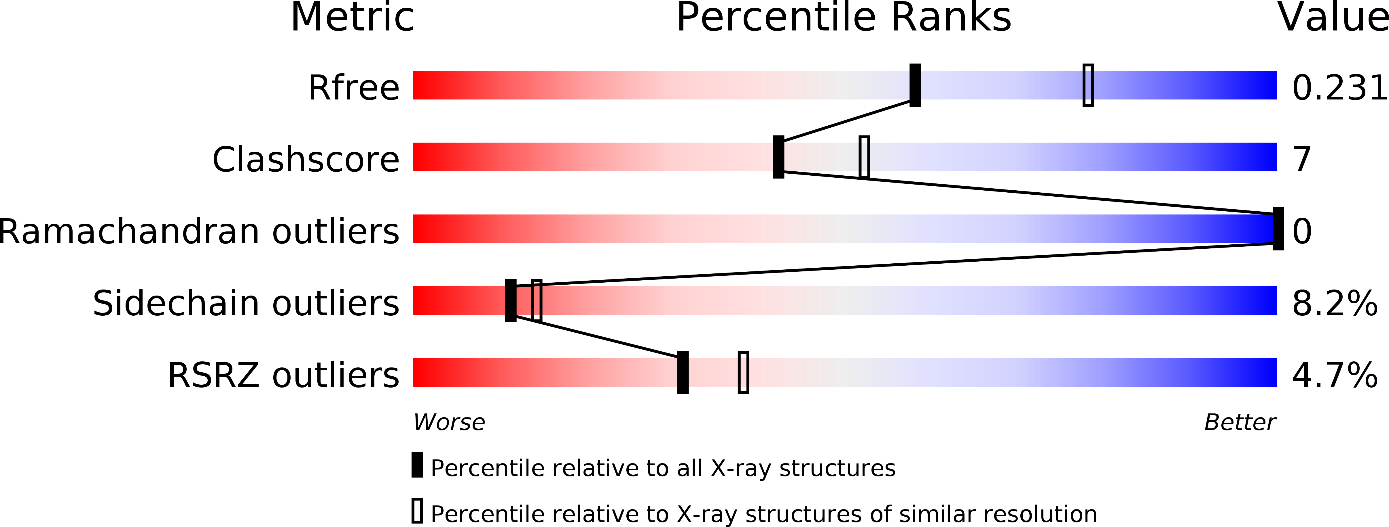
  • R-Value Free: 0.233 
  • R-Value Work: 0.181 
  • R-Value Observed: 0.183 

Starting Model: experimental
View more details

wwPDB Validation   3D Report Full Report


Ligand Structure Quality Assessment 


This is version 1.4 of the entry. See complete history


Literature

Crystal structure of the Rab9A-RUTBC2 RBD complex reveals the molecular basis for the binding specificity of Rab9A with RUTBC2.

Zhang, Z.Wang, S.Shen, T.Chen, J.Ding, J.

(2014) Structure 22: 1408-1420

  • DOI: https://doi.org/10.1016/j.str.2014.08.005
  • Primary Citation of Related Structures:  
    4QXA

  • PubMed Abstract: 

    Rab9 plays a vital role in regulating the transport of mannose 6-phosphate receptors from late endosomes to the trans-Golgi network through interactions with various effectors. Here, we report the crystal structure of GTP-bound Rab9A in complex with the Rab-binding domain (RBD) of the effector RUTBC2. RUTBC2 RBD assumes a pleckstrin homology domain fold that uses a binding site consisting of mainly β1 and the η1 insertion to interact with the switch and interswitch regions of Rab9A. The C-terminal hypervariable region of Rab9A is disordered and thus not required for RUTBC2 binding. The conformational plasticity of the switch and interswitch regions of Rab9A primarily determines the specificity for RUTBC2. Our biochemical and biological data confirm these findings and further show that Rab9B can bind to RUTBC2 probably in a similar manner as Rab9A. These results together reveal the molecular basis for the binding specificity of Rab9A with RUTBC2.


  • Organizational Affiliation

    State Key Laboratory of Molecular Biology, Institute of Biochemistry and Cell Biology, Shanghai Institutes for Biological Sciences, Chinese Academy of Sciences, 320 Yue-Yang Road, Shanghai 200031, China.


Macromolecules
Find similar proteins by:  (by identity cutoff)  |  3D Structure
Entity ID: 1
MoleculeChains Sequence LengthOrganismDetailsImage
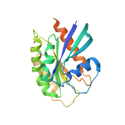
Ras-related protein Rab-9A208Mus musculusMutation(s): 1 
Gene Names: Rab9aRab9Sid99
UniProt
Find proteins for Q9R0M6 (Mus musculus)
Explore Q9R0M6 
Go to UniProtKB:  Q9R0M6
Entity Groups  
Sequence Clusters30% Identity50% Identity70% Identity90% Identity95% Identity100% Identity
UniProt GroupQ9R0M6
Sequence Annotations
Expand
  • Reference Sequence
Find similar proteins by:  (by identity cutoff)  |  3D Structure
Entity ID: 2
MoleculeChains Sequence LengthOrganismDetailsImage
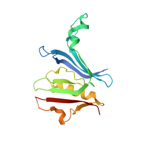
Small G protein signaling modulator 1180Mus musculusMutation(s): 0 
Gene Names: Sgsm1Rutbc2
UniProt & NIH Common Fund Data Resources
Find proteins for Q8BPQ7 (Mus musculus)
Explore Q8BPQ7 
Go to UniProtKB:  Q8BPQ7
IMPC:  MGI:107320
Entity Groups  
Sequence Clusters30% Identity50% Identity70% Identity90% Identity95% Identity100% Identity
UniProt GroupQ8BPQ7
Sequence Annotations
Expand
  • Reference Sequence
Experimental Data & Validation

Experimental Data

  • Method: X-RAY DIFFRACTION
  • Resolution: 2.30 Å
  • R-Value Free: 0.233 
  • R-Value Work: 0.181 
  • R-Value Observed: 0.183 
  • Space Group: P 21 21 21
Unit Cell:
Length ( Å )Angle ( ˚ )
a = 52.776α = 90
b = 59.944β = 90
c = 127.84γ = 90
Software Package:
Software NamePurpose
DENZOdata reduction
SCALEPACKdata scaling
PHASERphasing
PHENIXrefinement
PDB_EXTRACTdata extraction
HKL-2000data collection

Structure Validation

View Full Validation Report


Ligand Structure Quality Assessment 


Entry History 

Deposition Data

Revision History  (Full details and data files)

  • Version 1.0: 2014-09-10
    Type: Initial release
  • Version 1.1: 2014-09-24
    Changes: Database references
  • Version 1.2: 2017-11-22
    Changes: Refinement description
  • Version 1.3: 2019-12-25
    Changes: Database references
  • Version 1.4: 2023-11-08
    Changes: Data collection, Database references, Derived calculations, Refinement description