4OMB

Phosphate binding protein


Experimental Data Snapshot

  • Method: X-RAY DIFFRACTION
  • Resolution: 1.50 Å
  • R-Value Free: 0.216 
  • R-Value Work: 0.190 
  • R-Value Observed: 0.191 

wwPDB Validation   3D Report Full Report


This is version 1.3 of the entry. See complete history


Literature

The Pseudomonas aeruginosa phosphate transport protein PstS plays a phosphate-independent role in biofilm formation.

Neznansky, A.Blus-Kadosh, I.Yerushalmi, G.Banin, E.Opatowsky, Y.

(2014) FASEB J 28: 5223-5233

  • DOI: https://doi.org/10.1096/fj.14-258293
  • Primary Citation of Related Structures:  
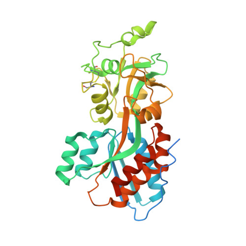
    4OMB

  • PubMed Abstract: 

    Pseudomonas aeruginosa (PA) is a primary cause of nosocomial infections. A key element in PA pathogenicity is its ability to form biofilms that withstand eradication by antibiotics and the immune system. Biofilm formation is controlled by phosphate signaling and here we provide evidence that PstS, a subunit of the PA Pst phosphate transporter, has a surprising role in this process. Using X-ray crystallography, we characterized the unique underpinnings of PstS phosphate binding and identified an unusual 15-residue N' loop extension. Structure-based experiments showed that PstS-mediated phosphate uptake and biofilm formation are in fact two distinct functions. Specifically, a point mutation that abrogated phosphate binding did not eliminate biofilm formation; conversely, truncation of the N' loop diminished the ability of PA to form biofilms but had no effect on phosphate binding and uptake. This places PstS at a junction that separately controls phosphate sensing and uptake and the ultrastructure organization of bacteria.


  • Organizational Affiliation

    Mina and Everard Goodman Faculty of Life Sciences and.


Macromolecules
Find similar proteins by:  (by identity cutoff)  |  3D Structure
Entity ID: 1
MoleculeChains Sequence LengthOrganismDetailsImage
Phosphate binding protein
A, B, C, D
347Pseudomonas aeruginosaMutation(s): 0 
Gene Names: pstS
Entity Groups  
Sequence Clusters30% Identity50% Identity70% Identity90% Identity95% Identity100% Identity
Sequence Annotations
Expand
  • Reference Sequence
Experimental Data & Validation

Experimental Data

  • Method: X-RAY DIFFRACTION
  • Resolution: 1.50 Å
  • R-Value Free: 0.216 
  • R-Value Work: 0.190 
  • R-Value Observed: 0.191 
  • Space Group: P 21 21 21
Unit Cell:
Length ( Å )Angle ( ˚ )
a = 35.444α = 90
b = 148.092β = 90
c = 216.452γ = 90
Software Package:
Software NamePurpose
EDNAdata collection
BALBESphasing
REFMACrefinement
XDSdata reduction
XDSdata scaling

Structure Validation

View Full Validation Report


Entry History 

Deposition Data

Revision History  (Full details and data files)

  • Version 1.0: 2014-12-10
    Type: Initial release
  • Version 1.1: 2014-12-17
    Changes: Database references
  • Version 1.2: 2017-11-22
    Changes: Refinement description
  • Version 1.3: 2023-09-20
    Changes: Data collection, Database references, Derived calculations, Refinement description