4KBM

Structure of the Mtb CarD/RNAP Beta subunit B1-B2 domains complex


Experimental Data Snapshot

  • Method: X-RAY DIFFRACTION
  • Resolution: 2.11 Å
  • R-Value Free: 0.239 
  • R-Value Work: 0.208 
  • R-Value Observed: 0.209 

wwPDB Validation   3D Report Full Report


This is version 1.3 of the entry. See complete history


Literature

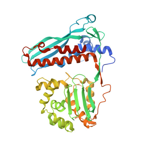
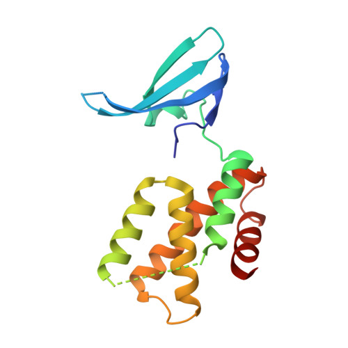
Structure of the Mtb CarD/RNAP beta-Lobes Complex Reveals the Molecular Basis of Interaction and Presents a Distinct DNA-Binding Domain for Mtb CarD.

Gulten, G.Sacchettini, J.C.

(2013) Structure 21: 1859-1869

  • DOI: https://doi.org/10.1016/j.str.2013.08.014
  • Primary Citation of Related Structures:  
    4KBJ, 4KBM

  • PubMed Abstract: 

    CarD from Mycobacterium tuberculosis (Mtb) is an essential protein shown to be involved in stringent response through downregulation of rRNA and ribosomal protein genes. CarD interacts with the β-subunit of RNAP and this interaction is vital for Mtb's survival during the persistent infection state. We have determined the crystal structure of CarD in complex with the RNAP β-subunit β1 and β2 domains at 2.1 Å resolution. The structure reveals the molecular basis of CarD/RNAP interaction, providing a basis to further our understanding of RNAP regulation by CarD. The structural fold of the CarD N-terminal domain is conserved in RNAP interacting proteins such as TRCF-RID and CdnL, and displays similar interactions to the predicted homology model based on the TRCF/RNAP β1 structure. Interestingly, the structure of the C-terminal domain, which is required for complete CarD function in vivo, represents a distinct DNA-binding fold.


  • Organizational Affiliation

    Department of Chemistry, Texas A&M University, College Station, TX 77843, USA.


Macromolecules
Find similar proteins by:  (by identity cutoff)  |  3D Structure
Entity ID: 1
MoleculeChains Sequence LengthOrganismDetailsImage
DNA-directed RNA polymerase subunit beta409Mycobacterium tuberculosisMutation(s): 0 
Gene Names: MT0695MTCI376.08crpoBRv0667
EC: 2.7.7.6
UniProt
Find proteins for P9WGY9 (Mycobacterium tuberculosis (strain ATCC 25618 / H37Rv))
Explore P9WGY9 
Go to UniProtKB:  P9WGY9
Entity Groups  
Sequence Clusters30% Identity50% Identity70% Identity90% Identity95% Identity100% Identity
UniProt GroupP9WGY9
Sequence Annotations
Expand
  • Reference Sequence
Find similar proteins by:  (by identity cutoff)  |  3D Structure
Entity ID: 2
MoleculeChains Sequence LengthOrganismDetailsImage
RNA polymerase-binding transcription factor CarD162Mycobacterium tuberculosisMutation(s): 0 
Gene Names: carDMT3689Rv3583c
UniProt
Find proteins for P9WJG3 (Mycobacterium tuberculosis (strain ATCC 25618 / H37Rv))
Explore P9WJG3 
Go to UniProtKB:  P9WJG3
Entity Groups  
Sequence Clusters30% Identity50% Identity70% Identity90% Identity95% Identity100% Identity
UniProt GroupP9WJG3
Sequence Annotations
Expand
  • Reference Sequence
Experimental Data & Validation

Experimental Data

  • Method: X-RAY DIFFRACTION
  • Resolution: 2.11 Å
  • R-Value Free: 0.239 
  • R-Value Work: 0.208 
  • R-Value Observed: 0.209 
  • Space Group: C 2 2 21
Unit Cell:
Length ( Å )Angle ( ˚ )
a = 49.152α = 90
b = 128.889β = 90
c = 225.526γ = 90
Software Package:
Software NamePurpose
HKL-2000data collection
PHENIXmodel building
PHENIXrefinement
HKL-2000data reduction
HKL-2000data scaling
PHENIXphasing

Structure Validation

View Full Validation Report


Entry History 

Deposition Data

Revision History  (Full details and data files)

  • Version 1.0: 2013-10-02
    Type: Initial release
  • Version 1.1: 2013-10-09
    Changes: Database references
  • Version 1.2: 2013-10-30
    Changes: Database references
  • Version 1.3: 2023-09-20
    Changes: Data collection, Database references, Refinement description