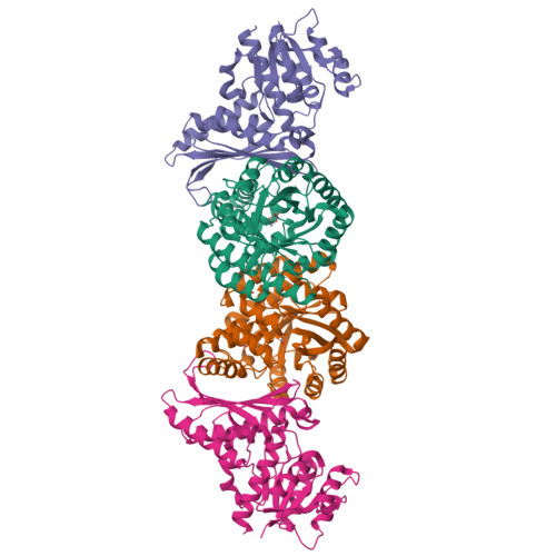
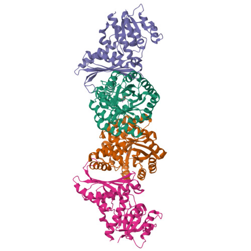
4JN6

Crystal Structure of the Aldolase-Dehydrogenase Complex from Mycobacterium tuberculosis HRv37


Experimental Data Snapshot

  • Method: X-RAY DIFFRACTION
  • Resolution: 1.93 Å
  • R-Value Free: 0.206 
  • R-Value Work: 0.174 
  • R-Value Observed: 0.176 

Starting Model: experimental
View more details

wwPDB Validation   3D Report Full Report


This is version 1.3 of the entry. See complete history


Literature

Characterization of an Aldolase-Dehydrogenase Complex from the Cholesterol Degradation Pathway of Mycobacterium tuberculosis.

Carere, J.McKenna, S.E.Kimber, M.S.Seah, S.Y.

(2013) Biochemistry 52: 3502-3511

  • DOI: https://doi.org/10.1021/bi400351h
  • Primary Citation of Related Structures:  
    4JN6

  • PubMed Abstract: 

    HsaF and HsaG are an aldolase and dehydrogenase from the cholesterol degradation pathway of Mycobacterium tuberculosis. HsaF could be heterologously expressed and purified as a soluble dimer, but the enzyme was inactive in the absence of HsaG. HsaF catalyzes the aldol cleavage of 4-hydroxy-2-oxoacids to produce pyruvate and an aldehyde. The enzyme requires divalent metals for activity, with a preference for Mn(2+). The Km values for 4-hydroxy-2-oxoacids were about 20-fold lower than observed for the aldolase homologue, BphI from the polychlorinated biphenyl degradation pathway. Acetaldehyde and propionaldehyde were channeled directly to the dehydrogenase, HsaG, without export to the bulk solvent where they were transformed to acyl-CoA in an NAD(+) and coenzyme A dependent reaction. HsaG is able to utilize aldehydes up to five carbons in length as substrates, with similar catalytic efficiencies. The HsaF-HsaG complex was crystallized and its structure was determined to a resolution of 1.93 Å. Substitution of serine 41 in HsaG with isoleucine or aspartate resulted in about 35-fold increase in Km for CoA but only 4-fold increase in Km dephospho-CoA, suggesting that this residue interacts with the 3'-ribose phosphate of CoA. A second protein annotated as a 4-hydroxy-2-oxopentanoic acid aldolase in M. tuberculosis (MhpE, Rv3469c) was expressed and purified, but was found to lack aldolase activity. Instead this enzyme was found to possess oxaloacetate decarboxylase activity, consistent with the conservation (with the 4-hydroxy-2-oxoacid aldolases) of residues involved in pyruvate enolate stabilization.


  • Organizational Affiliation

    Department of Molecular and Cellular Biology, University of Guelph , Guelph, Ontario, Canada N1G 2W1.


Macromolecules
Find similar proteins by:  (by identity cutoff)  |  3D Structure
Entity ID: 1
MoleculeChains Sequence LengthOrganismDetailsImage
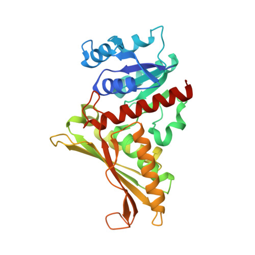
4-hydroxy-2-oxovalerate aldolase
A, C
346Mycobacterium tuberculosisMutation(s): 0 
Gene Names: Rv3534cMT3638
EC: 4.1.3.39 (PDB Primary Data), 4.1.3.43 (UniProt)
UniProt
Find proteins for P9WMK5 (Mycobacterium tuberculosis (strain ATCC 25618 / H37Rv))
Explore P9WMK5 
Go to UniProtKB:  P9WMK5
Entity Groups  
Sequence Clusters30% Identity50% Identity70% Identity90% Identity95% Identity100% Identity
UniProt GroupP9WMK5
Sequence Annotations
Expand
  • Reference Sequence
Find similar proteins by:  (by identity cutoff)  |  3D Structure
Entity ID: 2
MoleculeChains Sequence LengthOrganismDetailsImage
Acetaldehyde dehydrogenase
B, D
306Mycobacterium tuberculosisMutation(s): 0 
Gene Names: mhpFRv3535cMT3639
EC: 1.2.1.10 (PDB Primary Data), 1.2.1.87 (UniProt)
UniProt
Find proteins for P9WQH3 (Mycobacterium tuberculosis (strain ATCC 25618 / H37Rv))
Explore P9WQH3 
Go to UniProtKB:  P9WQH3
Entity Groups  
Sequence Clusters30% Identity50% Identity70% Identity90% Identity95% Identity100% Identity
UniProt GroupP9WQH3
Sequence Annotations
Expand
  • Reference Sequence
Experimental Data & Validation

Experimental Data

  • Method: X-RAY DIFFRACTION
  • Resolution: 1.93 Å
  • R-Value Free: 0.206 
  • R-Value Work: 0.174 
  • R-Value Observed: 0.176 
  • Space Group: C 1 2 1
Unit Cell:
Length ( Å )Angle ( ˚ )
a = 69.69α = 90
b = 142.69β = 95.08
c = 148.17γ = 90
Software Package:
Software NamePurpose
MxDXdata collection
PHENIXmodel building
PHENIXrefinement
XDSdata reduction
XSCALEdata scaling
PHENIXphasing

Structure Validation

View Full Validation Report


Entry History 

Deposition Data

Revision History  (Full details and data files)

  • Version 1.0: 2013-05-08
    Type: Initial release
  • Version 1.1: 2013-11-20
    Changes: Database references
  • Version 1.2: 2013-12-25
    Changes: Database references
  • Version 1.3: 2023-09-20
    Changes: Data collection, Database references, Derived calculations, Refinement description