4CKJ

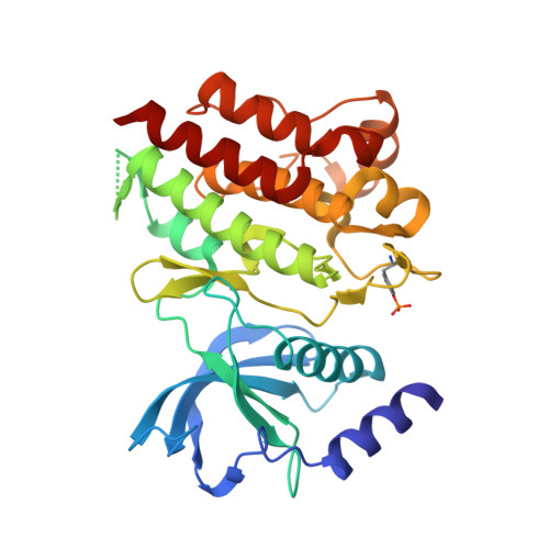
Crystal structure of RET tyrosine kinase domain bound to adenosine


Experimental Data Snapshot

  • Method: X-RAY DIFFRACTION
  • Resolution: 1.65 Å
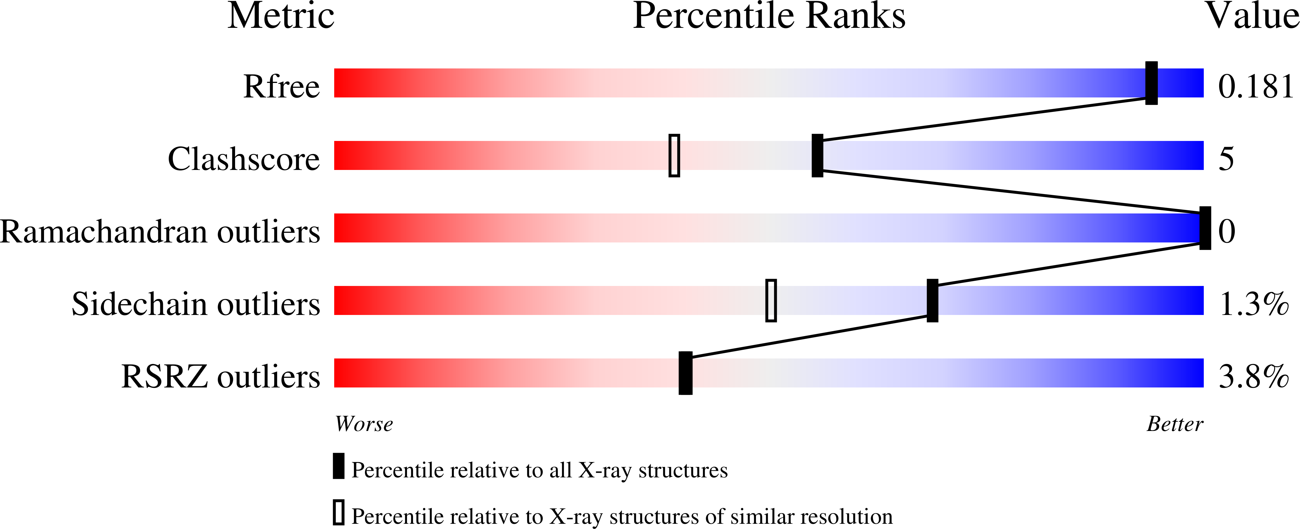
  • R-Value Free: 0.181 
  • R-Value Work: 0.153 
  • R-Value Observed: 0.155 

Starting Model: experimental
View more details

wwPDB Validation   3D Report Full Report


Ligand Structure Quality Assessment 


This is version 1.4 of the entry. See complete history


Literature

Oncogenic RET kinase domain mutations perturb the autophosphorylation trajectory by enhancing substrate presentation in trans.

Plaza-Menacho, I.Barnouin, K.Goodman, K.Martinez-Torres, R.J.Borg, A.Murray-Rust, J.Mouilleron, S.Knowles, P.McDonald, N.Q.

(2014) Mol Cell 53: 738-751

  • DOI: https://doi.org/10.1016/j.molcel.2014.01.015
  • Primary Citation of Related Structures:  
    4CKI, 4CKJ

  • PubMed Abstract: 

    To decipher the molecular basis for RET kinase activation and oncogenic deregulation, we defined the temporal sequence of RET autophosphorylation by label-free quantitative mass spectrometry. Early autophosphorylation sites map to regions flanking the kinase domain core, while sites within the activation loop only form at later time points. Comparison with oncogenic RET kinase revealed that late autophosphorylation sites become phosphorylated much earlier than wild-type RET, which is due to a combination of an enhanced enzymatic activity, increased ATP affinity, and surprisingly, by providing a better intermolecular substrate. Structural analysis of oncogenic M918T and wild-type RET kinase domains reveal a cis-inhibitory mechanism involving tethering contacts between the glycine-rich loop, activation loop, and αC-helix. Tether mutations only affected substrate presentation but perturbed the autophosphorylation trajectory similar to oncogenic mutations. This study reveals an unappreciated role for oncogenic RET kinase mutations in promoting intermolecular autophosphorylation by enhancing substrate presentation.


  • Organizational Affiliation

    Structural Biology Laboratory, London Research Institute, Cancer Research UK, WC2A 3LY London, UK. Electronic address: ivan.plaza-menacho@cancer.org.uk.


Macromolecules
Find similar proteins by:  (by identity cutoff)  |  3D Structure
Entity ID: 1
MoleculeChains Sequence LengthOrganismDetailsImage
PROTO-ONCOGENE TYROSINE-PROTEIN KINASE RECEPTOR RET314Homo sapiensMutation(s): 0 
EC: 2.7.10.1
UniProt & NIH Common Fund Data Resources
Find proteins for P07949 (Homo sapiens)
Explore P07949 
Go to UniProtKB:  P07949
PHAROS:  P07949
GTEx:  ENSG00000165731 
Entity Groups  
Sequence Clusters30% Identity50% Identity70% Identity90% Identity95% Identity100% Identity
UniProt GroupP07949
Sequence Annotations
Expand
  • Reference Sequence
Small Molecules
Ligands 2 Unique
IDChains Name / Formula / InChI Key2D Diagram3D Interactions
ADN
Query on ADN

Download Ideal Coordinates CCD File 
B [auth A]ADENOSINE
C10 H13 N5 O4
OIRDTQYFTABQOQ-KQYNXXCUSA-N
FMT
Query on FMT

Download Ideal Coordinates CCD File 
C [auth A]
D [auth A]
E [auth A]
F [auth A]
G [auth A]
C [auth A],
D [auth A],
E [auth A],
F [auth A],
G [auth A],
H [auth A],
I [auth A],
J [auth A],
K [auth A],
L [auth A],
M [auth A],
N [auth A],
O [auth A],
P [auth A],
Q [auth A],
R [auth A]
FORMIC ACID
C H2 O2
BDAGIHXWWSANSR-UHFFFAOYSA-N
Modified Residues  1 Unique
IDChains TypeFormula2D DiagramParent
PTR
Query on PTR
A
L-PEPTIDE LINKINGC9 H12 N O6 PTYR
Experimental Data & Validation

Experimental Data

  • Method: X-RAY DIFFRACTION
  • Resolution: 1.65 Å
  • R-Value Free: 0.181 
  • R-Value Work: 0.153 
  • R-Value Observed: 0.155 
  • Space Group: C 1 2 1
Unit Cell:
Length ( Å )Angle ( ˚ )
a = 73.19α = 90
b = 69.09β = 101.76
c = 78.64γ = 90
Software Package:
Software NamePurpose
PHENIXrefinement
PHASERphasing

Structure Validation

View Full Validation Report


Ligand Structure Quality Assessment 


Entry History 

Deposition Data

Revision History  (Full details and data files)

  • Version 1.0: 2014-03-05
    Type: Initial release
  • Version 1.1: 2014-03-19
    Changes: Database references, Other
  • Version 1.2: 2018-02-28
    Changes: Database references
  • Version 1.3: 2023-12-20
    Changes: Data collection, Database references, Derived calculations, Other, Refinement description
  • Version 1.4: 2024-11-13
    Changes: Structure summary