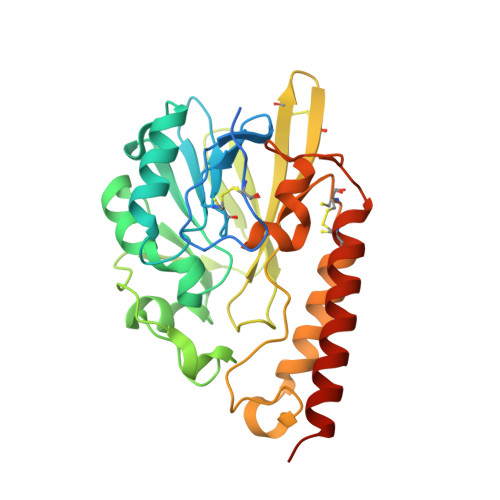
4AX1

Q157N mutant. Crystal Structure of the Mobile Metallo-beta-Lactamase AIM-1 from Pseudomonas aeruginosa: Insights into Antibiotic Binding and the role of Gln157


Experimental Data Snapshot

  • Method: X-RAY DIFFRACTION
  • Resolution: 1.40 Å
  • R-Value Free: 0.150 
  • R-Value Work: 0.130 
  • R-Value Observed: 0.131 

wwPDB Validation   3D Report Full Report


This is version 1.5 of the entry. See complete history


Literature

Crystal Structure of the Mobile Metallo-Beta-Lactamase Aim-1 from Pseudomonas Aeruginosa: Insights Into Antibiotic Binding and the Role of Gln157

Leiros, H.-K.S.Borra, P.S.Brandsdal, B.O.Edvardsen, K.S.W.Spencer, J.Walsh, T.R.Samuelsen, O.

(2012) Antimicrob Agents Chemother 56: 4341

  • DOI: https://doi.org/10.1128/AAC.00448-12
  • Primary Citation of Related Structures:  
    4AWY, 4AWZ, 4AX0, 4AX1

  • PubMed Abstract: 

    Metallo-β-lactamase (MBL) genes confer resistance to virtually all β-lactam antibiotics and are rapidly disseminated by mobile genetic elements in Gram-negative bacteria. MBLs belong to three different subgroups, B1, B2, and B3, with the mobile MBLs largely confined to subgroup B1. The B3 MBLs are a divergent subgroup of predominantly chromosomally encoded enzymes. AIM-1 (Adelaide IMipenmase 1) from Pseudomonas aeruginosa was the first B3 MBL to be identified on a readily mobile genetic element. Here we present the crystal structure of AIM-1 and use in silico docking and quantum mechanics and molecular mechanics (QM/MM) calculations, together with site-directed mutagenesis, to investigate its interaction with β-lactams. AIM-1 adopts the characteristic αβ/βα sandwich fold of MBLs but differs from other B3 enzymes in the conformation of an active site loop (residues 156 to 162) which is involved both in disulfide bond formation and, we suggest, interaction with substrates. The structure, together with docking and QM/MM calculations, indicates that the AIM-1 substrate binding site is narrower and more restricted than those of other B3 MBLs, possibly explaining its higher catalytic efficiency. The location of Gln157 adjacent to the AIM-1 zinc center suggests a role in drug binding that is supported by our in silico studies. However, replacement of this residue by either Asn or Ala resulted in only modest reductions in AIM-1 activity against the majority of β-lactam substrates, indicating that this function is nonessential. Our study reveals AIM-1 to be a subclass B3 MBL with novel structural and mechanistic features.


  • Organizational Affiliation

    The Norwegian Structural Biology Centre (NorStruct), Department of Chemistry, University of Tromsø, Tromsø, Norway. kirsti.leiros@uit.no


Macromolecules
Find similar proteins by:  (by identity cutoff)  |  3D Structure
Entity ID: 1
MoleculeChains Sequence LengthOrganismDetailsImage
METALLO-BETA-LACTAMASE AIM-1A [auth B]303Pseudomonas aeruginosaMutation(s): 1 
UniProt
Find proteins for B5DCA0 (Pseudomonas aeruginosa)
Explore B5DCA0 
Go to UniProtKB:  B5DCA0
Entity Groups  
Sequence Clusters30% Identity50% Identity70% Identity90% Identity95% Identity100% Identity
UniProt GroupB5DCA0
Sequence Annotations
Expand
  • Reference Sequence
Small Molecules
Ligands 4 Unique
IDChains Name / Formula / InChI Key2D Diagram3D Interactions
ZN
Query on ZN

Download Ideal Coordinates CCD File 
B,
C [auth B]
ZINC ION
Zn
PTFCDOFLOPIGGS-UHFFFAOYSA-N
ACT
Query on ACT

Download Ideal Coordinates CCD File 
G [auth B]ACETATE ION
C2 H3 O2
QTBSBXVTEAMEQO-UHFFFAOYSA-M
CA
Query on CA

Download Ideal Coordinates CCD File 
D [auth B],
H [auth B]
CALCIUM ION
Ca
BHPQYMZQTOCNFJ-UHFFFAOYSA-N
MG
Query on MG

Download Ideal Coordinates CCD File 
E [auth B],
F [auth B]
MAGNESIUM ION
Mg
JLVVSXFLKOJNIY-UHFFFAOYSA-N
Experimental Data & Validation

Experimental Data

  • Method: X-RAY DIFFRACTION
  • Resolution: 1.40 Å
  • R-Value Free: 0.150 
  • R-Value Work: 0.130 
  • R-Value Observed: 0.131 
  • Space Group: P 61 2 2
Unit Cell:
Length ( Å )Angle ( ˚ )
a = 77.992α = 90
b = 77.992β = 90
c = 239.766γ = 120
Software Package:
Software NamePurpose
REFMACrefinement
XDSdata reduction
XSCALEdata scaling

Structure Validation

View Full Validation Report


Entry History 

Deposition Data

Revision History  (Full details and data files)

  • Version 1.0: 2012-06-20
    Type: Initial release
  • Version 1.1: 2012-08-15
    Changes: Database references
  • Version 1.2: 2018-01-17
    Changes: Data collection
  • Version 1.3: 2018-06-20
    Changes: Data collection, Derived calculations
  • Version 1.4: 2019-10-16
    Changes: Data collection, Experimental preparation, Other
  • Version 1.5: 2024-10-09
    Changes: Data collection, Database references, Derived calculations, Structure summary