3WVF

Crystal structure of YidC from Escherichia coli


Experimental Data Snapshot

  • Method: X-RAY DIFFRACTION
  • Resolution: 3.20 Å
  • R-Value Free: 0.277 
  • R-Value Work: 0.221 
  • R-Value Observed: 0.227 

wwPDB Validation   3D Report Full Report


This is version 1.1 of the entry. See complete history


Literature

Crystal structure of Escherichia coli YidC, a membrane protein chaperone and insertase

Kumazaki, K.Kishimoto, T.Furukawa, A.Mori, H.Tanaka, Y.Dohmae, N.Ishitani, R.Tsukazaki, T.Nureki, O.

(2014) Sci Rep 4: 7299-7299

  • DOI: https://doi.org/10.1038/srep07299
  • Primary Citation of Related Structures:  
    3WVF

  • PubMed Abstract: 

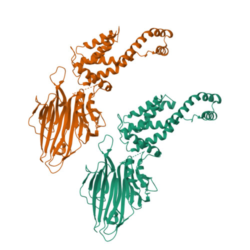
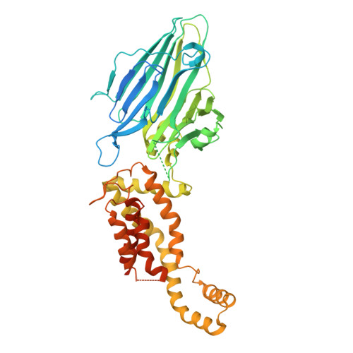
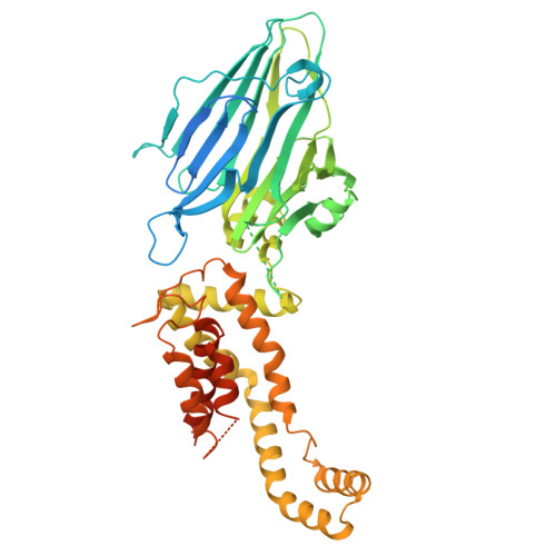
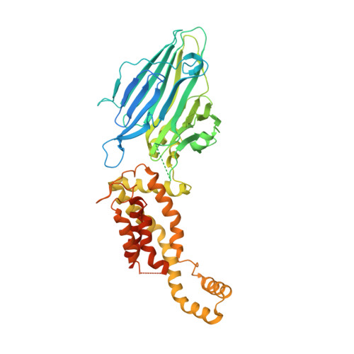
    Bacterial YidC, an evolutionally conserved membrane protein, functions as a membrane protein chaperone in cooperation with the Sec translocon and as an independent insertase for membrane proteins. In Gram-negative bacteria, the transmembrane and periplasmic regions of YidC interact with the Sec proteins, forming a multi-protein complex for Sec-dependent membrane protein integration. Here, we report the crystal structure of full-length Escherichia coli YidC. The structure reveals that a hydrophilic groove, formed by five transmembrane helices, is a conserved structural feature of YidC, as compared to the previous YidC structure from Bacillus halodurans, which lacks a periplasmic domain. Structural mapping of the substrate- or Sec protein-contact sites suggested the importance of the groove for the YidC functions as a chaperone and an insertase, and provided structural insight into the multi-protein complex.


  • Organizational Affiliation

    1] Department of Biological Sciences, Graduate School of Science, The University of Tokyo, 7-3-1 Hongo, Bunkyo-ku, Tokyo 113-0033, Japan [2] Global Research Cluster, RIKEN, 2-1 Hirosawa, Wako-shi, Saitama 351-0198, Japan.


Macromolecules
Find similar proteins by:  (by identity cutoff)  |  3D Structure
Entity ID: 1
MoleculeChains Sequence LengthOrganismDetailsImage
Membrane protein insertase YidC
A, B
551Escherichia coli UTI89Mutation(s): 0 
Gene Names: yidC
Membrane Entity: Yes 
UniProt
Find proteins for Q1R4M9 (Escherichia coli (strain UTI89 / UPEC))
Explore Q1R4M9 
Go to UniProtKB:  Q1R4M9
Entity Groups  
Sequence Clusters30% Identity50% Identity70% Identity90% Identity95% Identity100% Identity
UniProt GroupQ1R4M9
Sequence Annotations
Expand
  • Reference Sequence
Experimental Data & Validation

Experimental Data

  • Method: X-RAY DIFFRACTION
  • Resolution: 3.20 Å
  • R-Value Free: 0.277 
  • R-Value Work: 0.221 
  • R-Value Observed: 0.227 
  • Space Group: P 1
Unit Cell:
Length ( Å )Angle ( ˚ )
a = 42.729α = 78.08
b = 76.011β = 82.45
c = 91.318γ = 77.55
Software Package:
Software NamePurpose
HKL-2000data collection
PHENIXmodel building
PHENIXrefinement
HKL-2000data reduction
HKL-2000data scaling
PHENIXphasing

Structure Validation

View Full Validation Report


Entry History 

Deposition Data

Revision History  (Full details and data files)

  • Version 1.0: 2014-12-17
    Type: Initial release
  • Version 1.1: 2023-11-08
    Changes: Data collection, Database references, Refinement description