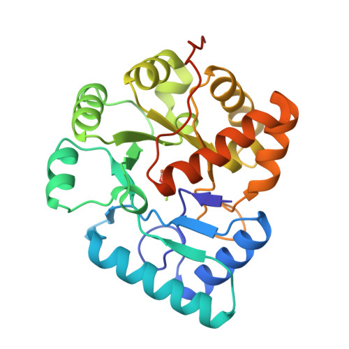
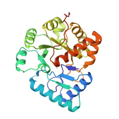
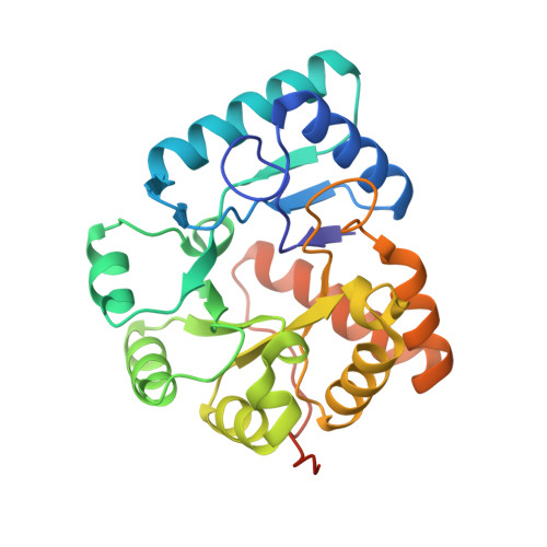
Crystal structures of YwqE from Bacillus subtilis and CpsB from Streptococcus pneumoniae, unique metal-dependent tyrosine phosphatases.
Kim, H.S., Lee, S.J., Yoon, H.J., An, D.R., Kim, D.J., Kim, S.J., Suh, S.W.(2011) J Struct Biol 175: 442-450
- PubMed: 21605684
- DOI: https://doi.org/10.1016/j.jsb.2011.05.007
- Primary Citation of Related Structures:
3QY6, 3QY7, 3QY8 - PubMed Abstract:
Unique metal-dependent protein tyrosine phosphatases that belong to the polymerase and histindinol phosphatase (PHP) family are present in Gram-positive bacteria. They are distinct from the Cys-based, low-molecular-weight phosphotyrosine protein phosphatases (LMPTPs). Two representative members of the PHP family tyrosine phosphatases are YwqE from Bacillus subtilis and CpsB from Streptococcus pneumoniae. YwqE is involved in polysaccharide biosynthesis, bacterial DNA metabolism, and DNA damage response in B. subtilis. CpsB regulates capsular polysaccharide biosynthesis via tyrosine dephosphorylation of CpsD, its cognate tyrosine kinase, in S. pneumoniae. To gain insights into the active site and possible conformational changes of the metal-dependent tyrosine phosphatases from Gram-positive bacteria, we have determined the crystal structures of B. subtilis YwqE (in both the apo form and the phosphate-bound form) and S. pneumoniae CpsB (in the sulfate-bound form). Comparisons of the three structures reveal conformational plasticity of two active site loops. Furthermore, in both structures of the phosphate-bound YwqE and the sulfate-bound CpsB, the phosphate (or sulfate) ion is bound to a cluster of three metal ions in the active site, thus providing insight into the pre-catalytic state.
Organizational Affiliation:
Department of Chemistry, College of Natural Sciences, Seoul National University, Seoul 151-742, South Korea.


