3QD5

Crystal structure of a putative ribose-5-phosphate isomerase from Coccidioides immitis solved by combined iodide ion SAD and MR


Experimental Data Snapshot

  • Method: X-RAY DIFFRACTION
  • Resolution: 1.90 Å
  • R-Value Free: 0.205 
  • R-Value Work: 0.164 
  • R-Value Observed: 0.166 

wwPDB Validation   3D Report Full Report


This is version 1.3 of the entry. See complete history


Literature

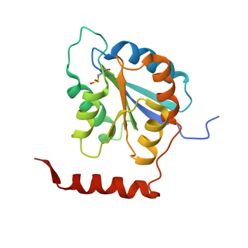
Structural characterization of a ribose-5-phosphate isomerase B from the pathogenic fungus Coccidioides immitis.

Edwards, T.E.Abramov, A.B.Smith, E.R.Baydo, R.O.Leonard, J.T.Leibly, D.J.Thompkins, K.B.Clifton, M.C.Gardberg, A.S.Staker, B.L.Van Voorhis, W.C.Myler, P.J.Stewart, L.J.

(2011) BMC Struct Biol 11: 39-39

  • DOI: https://doi.org/10.1186/1472-6807-11-39
  • Primary Citation of Related Structures:  
    3QD5, 3SDW, 3SGW

  • PubMed Abstract: 

    Ribose-5-phosphate isomerase is an enzyme that catalyzes the interconversion of ribose-5-phosphate and ribulose-5-phosphate. This family of enzymes naturally occurs in two distinct classes, RpiA and RpiB, which play an important role in the pentose phosphate pathway and nucleotide and co-factor biogenesis.


  • Organizational Affiliation

    Seattle Structural Genomics Center for Infectious Disease (SSGCID), USA. tedwards@embios.com


Macromolecules
Find similar proteins by:  (by identity cutoff)  |  3D Structure
Entity ID: 1
MoleculeChains Sequence LengthOrganismDetailsImage
putative ribose-5-phosphate isomerase
A, B
167Coccidioides immitis RSMutation(s): 0 
Gene Names: CIMG_079324559626
EC: 5.3.1.6
UniProt
Find proteins for P0CL19 (Coccidioides immitis (strain RS))
Explore P0CL19 
Go to UniProtKB:  P0CL19
Entity Groups  
Sequence Clusters30% Identity50% Identity70% Identity90% Identity95% Identity100% Identity
UniProt GroupP0CL19
Sequence Annotations
Expand
  • Reference Sequence
Small Molecules
Ligands 2 Unique
IDChains Name / Formula / InChI Key2D Diagram3D Interactions
IOD
Query on IOD

Download Ideal Coordinates CCD File 
AA [auth B]
BA [auth B]
CA [auth B]
D [auth A]
DA [auth B]
AA [auth B],
BA [auth B],
CA [auth B],
D [auth A],
DA [auth B],
E [auth A],
EA [auth B],
F [auth A],
FA [auth B],
G [auth A],
GA [auth B],
H [auth A],
I [auth A],
J [auth A],
K [auth A],
L [auth A],
M [auth A],
N [auth A],
O [auth A],
P [auth A],
R [auth B],
S [auth B],
T [auth B],
U [auth B],
V [auth B],
W [auth B],
X [auth B],
Y [auth B],
Z [auth B]
IODIDE ION
I
XMBWDFGMSWQBCA-UHFFFAOYSA-M
EDO
Query on EDO

Download Ideal Coordinates CCD File 
C [auth A],
Q [auth B]
1,2-ETHANEDIOL
C2 H6 O2
LYCAIKOWRPUZTN-UHFFFAOYSA-N
Modified Residues  1 Unique
IDChains TypeFormula2D DiagramParent
CSO
Query on CSO
A, B
L-PEPTIDE LINKINGC3 H7 N O3 SCYS
Experimental Data & Validation

Experimental Data

Unit Cell:
Length ( Å )Angle ( ˚ )
a = 103.15α = 90
b = 49.89β = 108.62
c = 61.97γ = 90
Software Package:
Software NamePurpose
XSCALEdata scaling
PHASERphasing
REFMACrefinement
PDB_EXTRACTdata extraction
StructureStudiodata collection
XDSdata reduction

Structure Validation

View Full Validation Report


Entry History 

Deposition Data

Revision History  (Full details and data files)

  • Version 1.0: 2011-02-09
    Type: Initial release
  • Version 1.1: 2011-07-13
    Changes: Version format compliance
  • Version 1.2: 2011-10-26
    Changes: Database references
  • Version 1.3: 2017-09-27
    Changes: Data collection