3HB9 | pdb_00003hb9

Crystal Structure of S. aureus Pyruvate Carboxylase A610T Mutant


Experimental Data Snapshot

  • Method: X-RAY DIFFRACTION
  • Resolution: 2.90 Å
  • R-Value Free: 
    0.266 (Depositor), 0.260 (DCC) 
  • R-Value Work: 
    0.220 (Depositor), 0.210 (DCC) 
  • R-Value Observed: 
    0.222 (Depositor) 

wwPDB Validation   3D Report Full Report


Ligand Structure Quality Assessment 

Created with Raphaël 2.3.0Worse 01 BetterLigand structure goodness of fit to experimental dataBest fitted ADPClick on this verticalbar to view detailsBest fitted BTIClick on this verticalbar to view details

This is version 1.3 of the entry. See complete history


Literature

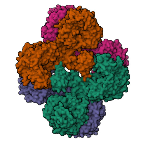
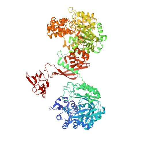
A Symmetrical Tetramer for S. aureus Pyruvate Carboxylase in Complex with Coenzyme A.

Yu, L.P.Xiang, S.Lasso, G.Gil, D.Valle, M.Tong, L.

(2009) Structure 17: 823-832

  • DOI: https://doi.org/10.1016/j.str.2009.04.008
  • Primary Citation of Related Structures:  
    3HB9, 3HBL, 3HO8

  • PubMed Abstract: 

    Pyruvate carboxylase (PC) is a conserved metabolic enzyme with important cellular functions. We report crystallographic and cryo-electron microscopy (EM) studies of Staphylococcus aureus PC (SaPC) in complex with acetyl-CoA, an allosteric activator, and mutagenesis, biochemical, and structural studies of the biotin binding site of its carboxyltransferase (CT) domain. The disease-causing A610T mutation abolishes catalytic activity by blocking biotin binding to the CT active site, and Thr908 might play a catalytic role in the CT reaction. The crystal structure of SaPC in complex with CoA reveals a symmetrical tetramer, with one CoA molecule bound to each monomer, and cryo-EM studies confirm the symmetrical nature of the tetramer. These observations are in sharp contrast to the highly asymmetrical tetramer of Rhizobium etli PC in complex with ethyl-CoA. Our structural information suggests that acetyl-CoA promotes a conformation for the dimer of the biotin carboxylase domain of PC that might be catalytically more competent.


  • Organizational Affiliation

    Department of Biological Sciences, Columbia University, New York, NY 10027, USA.


Macromolecules
Find similar proteins by:  (by identity cutoff)  |  3D Structure
Entity ID: 1
MoleculeChains Sequence LengthOrganismDetailsImage
Pyruvate carboxylase
A, B, C, D
1,150Staphylococcus aureus subsp. aureus Mu50Mutation(s): 1 
Gene Names: pycAPyruvate CarboxylaseSAV1114
EC: 6.4.1.1
UniProt
Find proteins for A0A0H3JRU9 (Staphylococcus aureus (strain Mu50 / ATCC 700699))
Explore A0A0H3JRU9 
Go to UniProtKB:  A0A0H3JRU9
Entity Groups  
Sequence Clusters30% Identity50% Identity70% Identity90% Identity95% Identity100% Identity
UniProt GroupA0A0H3JRU9
Sequence Annotations
Expand
  • Reference Sequence
Small Molecules
Ligands 3 Unique
IDChains Name / Formula / InChI Key2D Diagram3D Interactions
ADP
Query on ADP

Download Ideal Coordinates CCD File 
F [auth A],
K [auth C]
ADENOSINE-5'-DIPHOSPHATE
C10 H15 N5 O10 P2
XTWYTFMLZFPYCI-KQYNXXCUSA-N
BTI
Query on BTI

Download Ideal Coordinates CCD File 
E [auth A],
H [auth B],
J [auth C],
M [auth C]
5-(HEXAHYDRO-2-OXO-1H-THIENO[3,4-D]IMIDAZOL-6-YL)PENTANAL
C10 H16 N2 O2 S
ARDNWGMSCXSPBF-CIUDSAMLSA-N
MN
Query on MN

Download Ideal Coordinates CCD File 
G [auth A],
I [auth B],
L [auth C],
N [auth D]
MANGANESE (II) ION
Mn
WAEMQWOKJMHJLA-UHFFFAOYSA-N
Experimental Data & Validation

Experimental Data

  • Method: X-RAY DIFFRACTION
  • Resolution: 2.90 Å
  • R-Value Free:  0.266 (Depositor), 0.260 (DCC) 
  • R-Value Work:  0.220 (Depositor), 0.210 (DCC) 
  • R-Value Observed: 0.222 (Depositor) 
Space Group: P 1 21 1
Unit Cell:
Length ( Å )Angle ( ˚ )
a = 96.566α = 90
b = 256.763β = 109.65
c = 126.485γ = 90
Software Package:
Software NamePurpose
DENZOdata reduction
SCALEPACKdata scaling
COMOphasing
REFMACrefinement
PDB_EXTRACTdata extraction

Structure Validation

View Full Validation Report


Ligand Structure Quality Assessment 

Created with Raphaël 2.3.0Worse 01 BetterLigand structure goodness of fit to experimental dataBest fitted ADPClick on this verticalbar to view detailsBest fitted BTIClick on this verticalbar to view details

Entry History 

Deposition Data

Revision History  (Full details and data files)

  • Version 1.0: 2009-06-30
    Type: Initial release
  • Version 1.1: 2011-07-13
    Changes: Version format compliance
  • Version 1.2: 2017-11-01
    Changes: Refinement description
  • Version 1.3: 2021-10-13
    Changes: Database references, Derived calculations