3DC8

Crystal structure of dihydropyrimidinase from Sinorhizobium meliloti


Experimental Data Snapshot

  • Method: X-RAY DIFFRACTION
  • Resolution: 1.85 Å
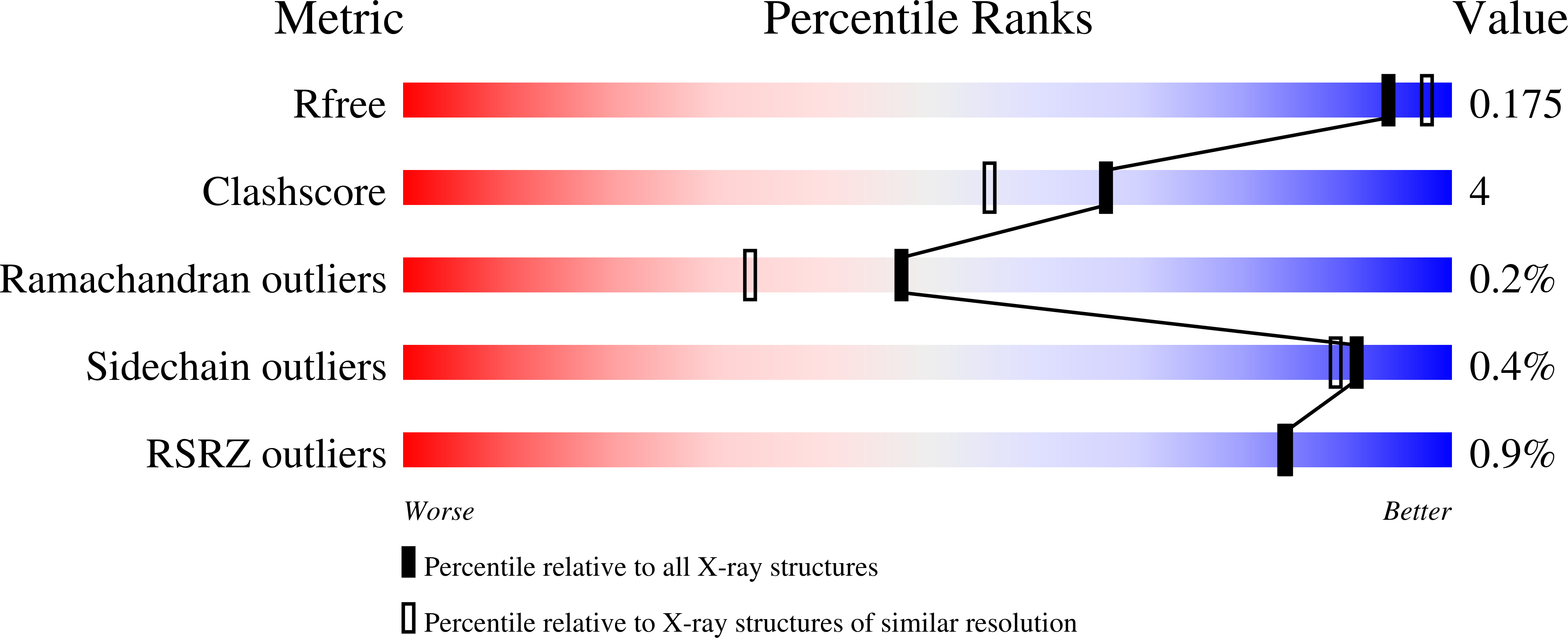
  • R-Value Free: 0.176 
  • R-Value Work: 0.148 
  • R-Value Observed: 0.149 

Starting Models: experimental
View more details

wwPDB Validation   3D Report Full Report


This is version 1.4 of the entry. See complete history


Literature

Structure of dihydropyrimidinase from Sinorhizobium meliloti CECT4114: new features in an amidohydrolase family member

Martinez-Rodriguez, S.Martinez-Gomez, A.I.Clemente-Jimenez, J.M.Rodriguez-Vico, F.Garcia-Ruiz, J.M.Las Heras-Vazquez, F.J.Gavira, J.A.

(2010) J Struct Biol 169: 200-208

  • DOI: https://doi.org/10.1016/j.jsb.2009.10.013
  • Primary Citation of Related Structures:  
    3DC8

  • PubMed Abstract: 

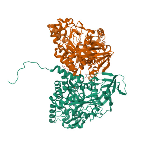
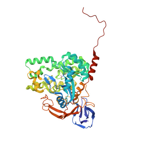
    The recombinant dihydropyrimidinase from Sinorhizobium meliloti CECT4114 (SmelDhp) has been characterised and its crystal structure elucidated at 1.85A. The global architecture of the protein is reminiscent of that of the amidohydrolase superfamily, consisting of two domains; an (alpha/beta)(8) TIM-like barrel domain, where the catalytic centre is located, and a smaller beta-sheet sandwich domain of unknown function. The c-terminal tails of each subunit extend toward another monomer in a swapping-like manner, creating a hydrogen bond network which suggests its implication in protein oligomerisation. Mutational and structural evidence suggest the involvement of a conserved tyrosine in the reaction mechanism of the enzyme. SmelDhp presents both hydantoinase and dihydropyrimidinase activities, with higher affinity for the natural six-membered ring substrates. For the five-membered ring substrates, affinity was greater for those with aliphatic and apolar groups in the 5th carbon atom, with the highest rates of hydrolysis for d-5-methyl and d-5-ethyl hydantoin (k(cat)/K(m)=2736+/-380 and 944+/-52M(-1)s(-1), respectively). The optimal conditions for the enzyme activity were found to be 60 degrees C of temperature at pH 8.0. SmelDhp retains 95% of its activity after 6-hour preincubation at 60 degrees C. This is the first dihydropyrimidinase used for the hydrolytic opening of non-natural 6-monosubstituted dihydrouracils, which may be exploited for the production of beta-amino acids.


  • Organizational Affiliation

    Dpto. Química Física, Bioquímica y Química Inorgánica, Universidad de Almería, Almería, Spain. srodrig@ual.es


Macromolecules
Find similar proteins by:  (by identity cutoff)  |  3D Structure
Entity ID: 1
MoleculeChains Sequence LengthOrganismDetailsImage
Dihydropyrimidinase
A, B
490Sinorhizobium melilotiMutation(s): 0 
Gene Names: dhp
EC: 3.5.2.2
UniProt
Find proteins for Q0PQZ5 (Rhizobium meliloti)
Explore Q0PQZ5 
Go to UniProtKB:  Q0PQZ5
Entity Groups  
Sequence Clusters30% Identity50% Identity70% Identity90% Identity95% Identity100% Identity
UniProt GroupQ0PQZ5
Sequence Annotations
Expand
  • Reference Sequence
Small Molecules
Ligands 3 Unique
IDChains Name / Formula / InChI Key2D Diagram3D Interactions
GOL
Query on GOL

Download Ideal Coordinates CCD File 
F [auth A],
G [auth A],
H [auth A],
L [auth B]
GLYCEROL
C3 H8 O3
PEDCQBHIVMGVHV-UHFFFAOYSA-N
ZN
Query on ZN

Download Ideal Coordinates CCD File 
C [auth A],
D [auth A],
I [auth B],
J [auth B]
ZINC ION
Zn
PTFCDOFLOPIGGS-UHFFFAOYSA-N
ACT
Query on ACT

Download Ideal Coordinates CCD File 
E [auth A],
K [auth B]
ACETATE ION
C2 H3 O2
QTBSBXVTEAMEQO-UHFFFAOYSA-M
Modified Residues  1 Unique
IDChains TypeFormula2D DiagramParent
KCX
Query on KCX
A, B
L-PEPTIDE LINKINGC7 H14 N2 O4LYS
Experimental Data & Validation

Experimental Data

  • Method: X-RAY DIFFRACTION
  • Resolution: 1.85 Å
  • R-Value Free: 0.176 
  • R-Value Work: 0.148 
  • R-Value Observed: 0.149 
  • Space Group: C 2 2 21
Unit Cell:
Length ( Å )Angle ( ˚ )
a = 124.887α = 90
b = 126.281β = 90
c = 196.104γ = 90
Software Package:
Software NamePurpose
SAINTdata scaling
MOLREPphasing
REFMACrefinement
PDB_EXTRACTdata extraction
SAINTdata reduction

Structure Validation

View Full Validation Report


Entry History 

Deposition Data

Revision History  (Full details and data files)

  • Version 1.0: 2009-04-21
    Type: Initial release
  • Version 1.1: 2011-07-13
    Changes: Advisory, Version format compliance
  • Version 1.2: 2011-08-10
    Changes: Database references
  • Version 1.3: 2023-11-01
    Changes: Data collection, Database references, Derived calculations, Refinement description
  • Version 1.4: 2023-11-15
    Changes: Data collection