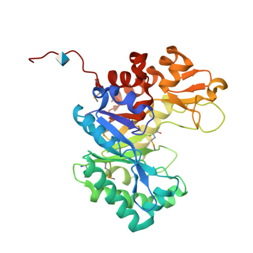
Crystal structure of a chitinase from Bacteroides thetaiotaomicron.
Damodharan, L., Burley, S.K., Swaminathan, S.To be published.
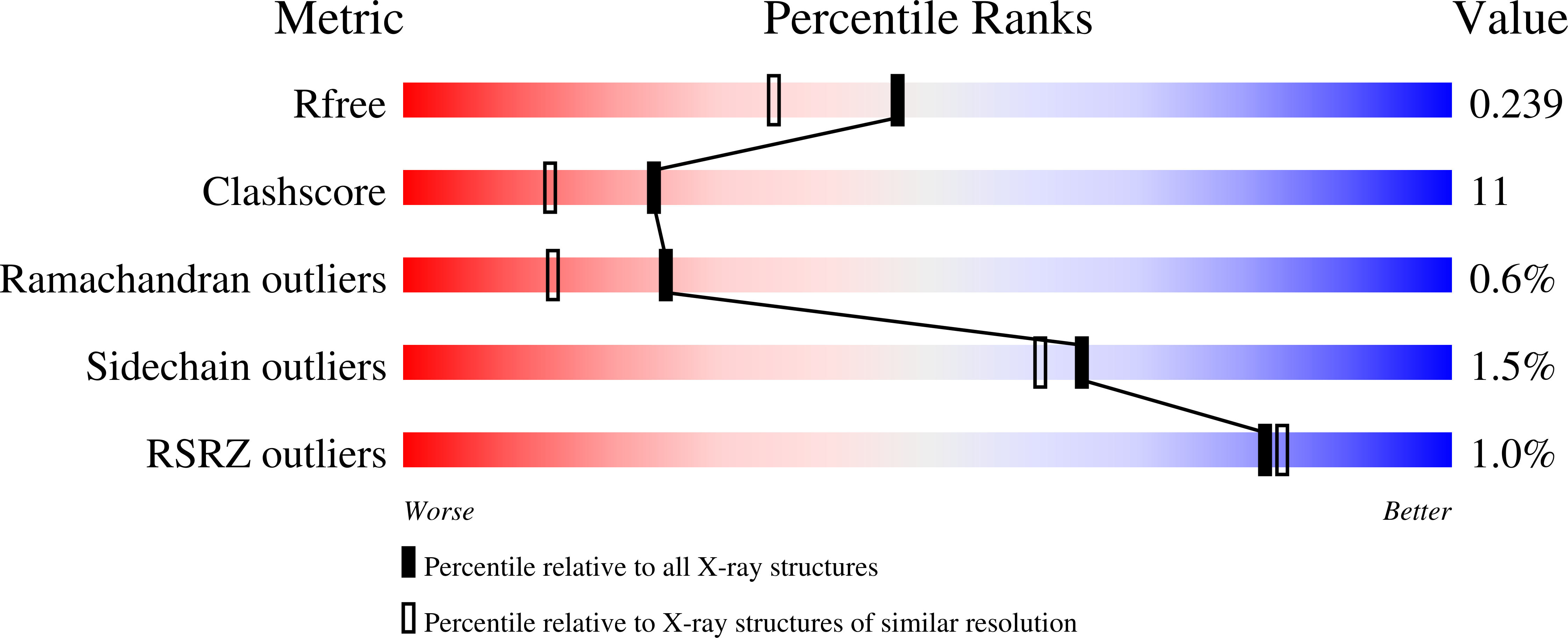
Experimental Data Snapshot
Entity ID: 1 | |||||
|---|---|---|---|---|---|
| Molecule | Chains | Sequence Length | Organism | Details | Image |
| Chitinase | 312 | Bacteroides thetaiotaomicron VPI-5482 | Mutation(s): 0 Gene Names: BT_2825 EC: 3.2.1.14 | 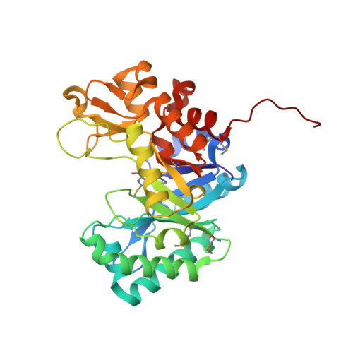 | |
UniProt | |||||
Find proteins for Q8A3X9 (Bacteroides thetaiotaomicron (strain ATCC 29148 / DSM 2079 / JCM 5827 / CCUG 10774 / NCTC 10582 / VPI-5482 / E50)) Explore Q8A3X9 Go to UniProtKB: Q8A3X9 | |||||
Entity Groups | |||||
| Sequence Clusters | 30% Identity50% Identity70% Identity90% Identity95% Identity100% Identity | ||||
| UniProt Group | Q8A3X9 | ||||
Sequence AnnotationsExpand | |||||
| |||||
| Ligands 1 Unique | |||||
|---|---|---|---|---|---|
| ID | Chains | Name / Formula / InChI Key | 2D Diagram | 3D Interactions | |
| GCS Query on GCS | B [auth A] | 2-amino-2-deoxy-beta-D-glucopyranose C6 H13 N O5 MSWZFWKMSRAUBD-QZABAPFNSA-N |  | ||
| Modified Residues 1 Unique | |||||
|---|---|---|---|---|---|
| ID | Chains | Type | Formula | 2D Diagram | Parent |
| MSE Query on MSE | A | L-PEPTIDE LINKING | C5 H11 N O2 Se |  | MET |
| Length ( Å ) | Angle ( ˚ ) |
|---|---|
| a = 60.884 | α = 90 |
| b = 70.389 | β = 90 |
| c = 79.259 | γ = 90 |
| Software Name | Purpose |
|---|---|
| CNS | refinement |
| CBASS | data collection |
| HKL-2000 | data reduction |
| HKL-2000 | data scaling |
| SHELXD | phasing |
| SHARP | phasing |
| ARP/wARP | model building |