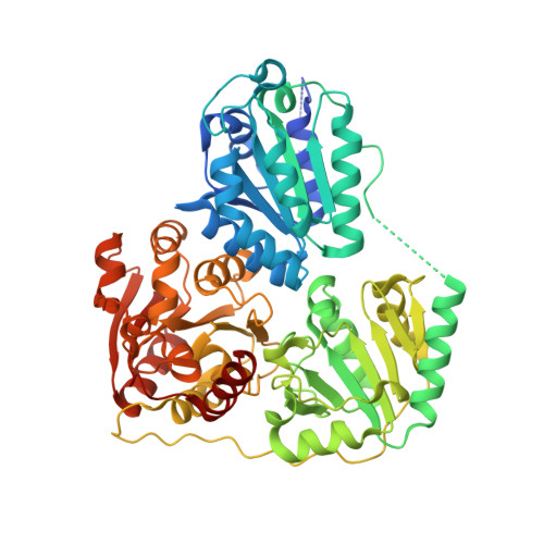
2VBG

The complex structure of the branched-chain keto acid decarboxylase (KdcA) from Lactococcus lactis with 2R-1-hydroxyethyl-deazaThDP


Experimental Data Snapshot

  • Method: X-RAY DIFFRACTION
  • Resolution: 1.80 Å
  • R-Value Free: 0.205 
  • R-Value Work: 0.163 
  • R-Value Observed: 0.165 

Starting Model: experimental
View more details

wwPDB Validation   3D Report Full Report


This is version 1.3 of the entry. See complete history


Literature

Crystal Structure of the Branched-Chain Keto Acid Decarboxylase (Kdca) from Lactococcus Lactis Provides Insights Into the Structural Basis for the Chemo- and Enantioselective Carboligation Reaction

Berthold, C.L.Gocke, D.Wood, M.D.Leeper, F.Pohl, M.Schneider, G.

(2007) Acta Crystallogr D Biol Crystallogr 63: 1217

  • DOI: https://doi.org/10.1107/S0907444907050433

  • PubMed Abstract: 

    The thiamin diphosphate (ThDP) dependent branched-chain keto acid decarboxylase (KdcA) from Lactococcus lactis catalyzes the decarboxylation of 3-methyl-2-oxobutanoic acid to 3-methylpropanal (isobutyraldehyde) and CO2. The enzyme is also able to catalyze carboligation reactions with an exceptionally broad substrate range, a feature that makes KdcA a potentially valuable biocatalyst for C-C bond formation, in particular for the enzymatic synthesis of diversely substituted 2-hydroxyketones with high enantioselectivity. The crystal structures of recombinant holo-KdcA and of a complex with an inhibitory ThDP analogue mimicking a reaction intermediate have been determined to resolutions of 1.6 and 1.8 A, respectively. KdcA shows the fold and cofactor-protein interactions typical of thiamin-dependent enzymes. In contrast to the tetrameric assembly displayed by most other ThDP-dependent decarboxylases of known structure, KdcA is a homodimer. The crystal structures provide insights into the structural basis of substrate selectivity and stereoselectivity of the enzyme and thus are suitable as a framework for the redesign of the substrate profile in carboligation reactions.


  • Organizational Affiliation

    Department of Medical Biochemistry and Biophysics, Karolinska Institutet, S-17177 Stockholm, Sweden.


Macromolecules
Find similar proteins by:  (by identity cutoff)  |  3D Structure
Entity ID: 1
MoleculeChains Sequence LengthOrganismDetailsImage
BRANCHED-CHAIN ALPHA-KETOACID DECARBOXYLASE
A, B
570Lactococcus lactisMutation(s): 0 
UniProt
Find proteins for Q6QBS4 (Lactococcus lactis)
Explore Q6QBS4 
Go to UniProtKB:  Q6QBS4
Entity Groups  
Sequence Clusters30% Identity50% Identity70% Identity90% Identity95% Identity100% Identity
UniProt GroupQ6QBS4
Sequence Annotations
Expand
  • Reference Sequence
Small Molecules
Ligands 2 Unique
IDChains Name / Formula / InChI Key2D Diagram3D Interactions
R1T
Query on R1T

Download Ideal Coordinates CCD File 
D [auth A],
F [auth B]
2-{4-[(4-AMINO-2-METHYLPYRIMIDIN-5-YL)METHYL]-5-[(1R)-1-HYDROXYETHYL]-3-METHYL-2-THIENYL}ETHYL TRIHYDROGEN DIPHOSPHATE
C15 H23 N3 O8 P2 S
ORVRYSKZCUVOLA-SECBINFHSA-N
MG
Query on MG

Download Ideal Coordinates CCD File 
C [auth A],
E [auth B]
MAGNESIUM ION
Mg
JLVVSXFLKOJNIY-UHFFFAOYSA-N
Experimental Data & Validation

Experimental Data

  • Method: X-RAY DIFFRACTION
  • Resolution: 1.80 Å
  • R-Value Free: 0.205 
  • R-Value Work: 0.163 
  • R-Value Observed: 0.165 
  • Space Group: P 21 21 21
Unit Cell:
Length ( Å )Angle ( ˚ )
a = 65.977α = 90
b = 108.578β = 90
c = 146.529γ = 90
Software Package:
Software NamePurpose
REFMACrefinement
MOSFLMdata reduction
SCALAdata scaling
MOLREPphasing

Structure Validation

View Full Validation Report


Entry History 

Deposition Data

Revision History  (Full details and data files)

  • Version 1.0: 2007-11-27
    Type: Initial release
  • Version 1.1: 2011-05-08
    Changes: Version format compliance
  • Version 1.2: 2011-07-13
    Changes: Version format compliance
  • Version 1.3: 2023-12-13
    Changes: Data collection, Database references, Derived calculations, Other, Refinement description