Structure, substrate recognition and reactivity of Leishmania major mevalonate kinase.
Sgraja, T., Smith, T.K., Hunter, W.N.(2007) BMC Struct Biol 7: 20-20
- PubMed: 17397541
- DOI: https://doi.org/10.1186/1472-6807-7-20
- Primary Citation of Related Structures:
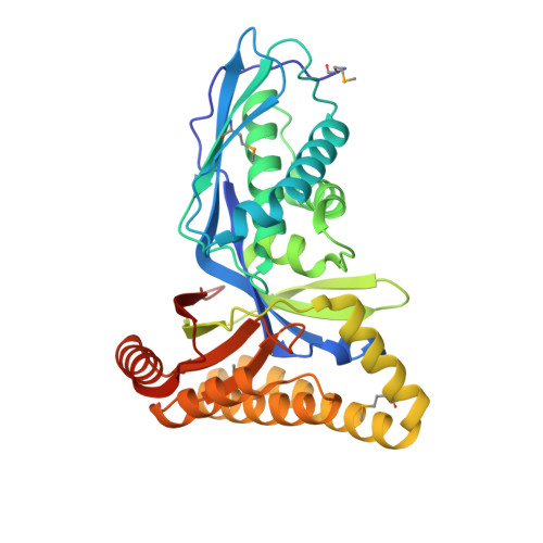
2HFS, 2HFU - PubMed Abstract:
Isoprenoid precursor synthesis via the mevalonate route in humans and pathogenic trypanosomatids is an important metabolic pathway. There is however, only limited information available on the structure and reactivity of the component enzymes in trypanosomatids. Since isoprenoid biosynthesis is essential for trypanosomatid viability and may provide new targets for therapeutic intervention it is important to characterize the pathway components. Putative mevalonate kinase encoding genes from Leishmania major (LmMK) and Trypanosoma brucei (TbMK) have been cloned, over-expressed in and proteins isolated from procyclic-form T. brucei. A highly sensitive radioactive assay was developed and shows ATP-dependent phosphorylation of mevalonate. Apo and (R)-mevalonate bound crystal structures of LmMK, from a bacterial expression system, have been determined to high resolution providing, for the first time, information concerning binding of mevalonate to an MK. The mevalonate binds in a deep cavity lined by highly conserved residues. His25 is key for binding and for discrimination of (R)- over (S)-mevalonate, with the main chain amide interacting with the C3 hydroxyl group of (R)-mevalonate, and the side chain contributing, together with Val202 and Thr283, to the construction of a hydrophobic binding site for the C3 methyl substituent. The C5 hydroxyl, where phosphorylation occurs, points towards catalytic residues, Lys18 and Asp155. The activity of LmMK was significantly reduced compared to MK from other species and we were unable to obtain ATP-binding data. Comparisons with the rat MK:ATP complex were used to investigate how this substrate might bind. In LmMK, helix alpha2 and the preceding polypeptide adopt a conformation, not seen in related kinase structures, impeding access to the nucleotide triphosphate binding site suggesting that a conformational rearrangement is required to allow ATP binding. Our new structural information, consistent with data on homologous enzymes allows a detailed description of how mevalonate is recognized and positioned for catalysis in MK. The mevalonate-binding site is highly conserved yet the ATP-binding site is structurally distinct in LmMK. We are unable to provide a definitive explanation for the low activity of recombinant protein isolated from a bacterial expression system compared to material isolated from procyclic-form Trypanosoma brucei.
Organizational Affiliation:
Division of Biological Chemistry and Molecular Microbiology, School of Life Sciences, University of Dundee, Dundee, UK. tanja.sgraja@web.de