2BS9

Native crystal structure of a GH39 beta-xylosidase XynB1 from Geobacillus stearothermophilus


Experimental Data Snapshot

  • Method: X-RAY DIFFRACTION
  • Resolution: 2.20 Å
  • R-Value Free: 0.259 
  • R-Value Work: 0.209 
  • R-Value Observed: 0.211 

wwPDB Validation   3D Report Full Report


This is version 1.2 of the entry. See complete history


Literature

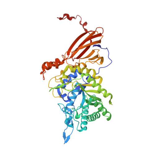
Enzyme-Substrate Complex Structures of a Gh39 Beta-Xylosidase from Geobacillus Stearothermophilus.

Czjzek, M.David, A.B.Bravman, T.Shoham, G.Henrissat, B.Shoham, Y.

(2005) J Mol Biol 353: 838

  • DOI: https://doi.org/10.1016/j.jmb.2005.09.003
  • Primary Citation of Related Structures:  
    2BFG, 2BS9

  • PubMed Abstract: 

    Beta-D-Xylosidases are glycoside hydrolases that catalyse the release of xylose units from short xylooligosaccharides and are engaged in the final breakdown of plant cell-wall hemicelluloses. beta-D-Xylosidases are found in glycoside hydrolase families 3, 39, 43, 52 and 54. The first crystal structure of a GH39 beta-xylosidase revealed a multi-domain organization with the catalytic domain having the canonical (beta/alpha)8 barrel fold. Here, we report the crystal structure of the GH39 Geobacillus stearothermophilus beta-D-xylosidase, inactivated by a point mutation of the general acid-base residue E160A, in complex with the chromogenic substrate molecule 2,5-dinitrophenyl-beta-D-xyloside. Surprisingly, six of the eight active sites present in the crystallographic asymmetric unit contain the trapped covalent glycosyl-enzyme intermediate, while two of them still contain the uncleaved substrate. The structural characterization of these two critical species along the reaction coordinate of this enzyme identifies the residues forming its xyloside-binding pocket as well as those essential for its aglycone recognition.


  • Organizational Affiliation

    Station Biologique de Roscoff, Végétaux Marins et Biomolécules, UMR7139-CNRS-UPMC, Place George Teissier, BP74, 29682 Roscoff, France. czjzek@sb-roscoff.fr


Macromolecules
Find similar proteins by:  (by identity cutoff)  |  3D Structure
Entity ID: 1
MoleculeChains Sequence LengthOrganismDetailsImage
BETA-XYLOSIDASE
A, B, C, D, E
A, B, C, D, E, F, G, H
503Geobacillus stearothermophilusMutation(s): 0 
EC: 3.2.1.37
UniProt
Find proteins for Q9ZFM2 (Geobacillus stearothermophilus)
Explore Q9ZFM2 
Go to UniProtKB:  Q9ZFM2
Entity Groups  
Sequence Clusters30% Identity50% Identity70% Identity90% Identity95% Identity100% Identity
UniProt GroupQ9ZFM2
Sequence Annotations
Expand
  • Reference Sequence
Experimental Data & Validation

Experimental Data

  • Method: X-RAY DIFFRACTION
  • Resolution: 2.20 Å
  • R-Value Free: 0.259 
  • R-Value Work: 0.209 
  • R-Value Observed: 0.211 
  • Space Group: P 21 21 21
Unit Cell:
Length ( Å )Angle ( ˚ )
a = 92.668α = 90
b = 165.744β = 90
c = 311.004γ = 90
Software Package:
Software NamePurpose
REFMACrefinement
MOSFLMdata reduction
SCALAdata scaling
AMoREphasing

Structure Validation

View Full Validation Report


Entry History 

Deposition Data

Revision History  (Full details and data files)

  • Version 1.0: 2005-10-12
    Type: Initial release
  • Version 1.1: 2012-11-14
    Changes: Database references, Derived calculations, Non-polymer description, Other, Source and taxonomy, Structure summary, Version format compliance
  • Version 1.2: 2023-12-13
    Changes: Data collection, Database references, Derived calculations, Other, Refinement description