2BQ1

Ribonucleotide reductase class 1b holocomplex R1E,R2F from Salmonella typhimurium


Experimental Data Snapshot

  • Method: X-RAY DIFFRACTION
  • Resolution: 3.99 Å
  • R-Value Free: 0.310 
  • R-Value Work: 0.262 
  • R-Value Observed: 0.265 

wwPDB Validation   3D Report Full Report


Ligand Structure Quality Assessment 


This is version 1.2 of the entry. See complete history


Literature

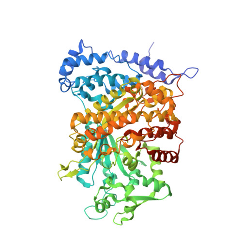
The First Holocomplex Structure of Ribonucleotide Reductase Gives New Insight Into its Mechanism of Action

Uppsten, M.Farnegardh, M.Domkin, V.Uhlin, U.

(2006) J Mol Biol 359: 365

  • DOI: https://doi.org/10.1016/j.jmb.2006.03.035
  • Primary Citation of Related Structures:  
    2BQ1

  • PubMed Abstract: 

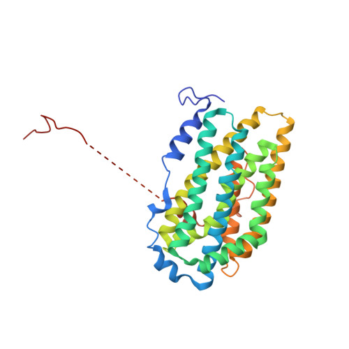
    Ribonucleotide reductase is an indispensable enzyme for all cells, since it catalyses the biosynthesis of the precursors necessary for both building and repairing DNA. The ribonucleotide reductase class I enzymes, present in all mammals as well as in many prokaryotes and DNA viruses, are composed mostly of two homodimeric proteins, R1 and R2. The reaction involves long-range radical transfer between the two proteins. Here, we present the first crystal structure of a ribonucleotide reductase R1/R2 holocomplex. The biological relevance of this complex is based on the binding of the R2 C terminus in the hydrophobic cleft of R1, an interaction proven to be crucial for enzyme activity, and by the fact that all conserved amino acid residues in R2 are facing the R1 active sites. We suggest that the asymmetric R1/R2 complex observed in the 4A crystal structure of Salmonella typhimurium ribonucleotide reductase represents an intermediate stage in the reaction cycle, and at the moment of reaction the homodimers transiently form a tight symmetric complex.


  • Organizational Affiliation

    Department of Molecular Biology, Swedish University of Agricultural Sciences, Uppsala Biomedical Center, Box 590, SE-751 24 Uppsala, Sweden.


Macromolecules
Find similar proteins by:  (by identity cutoff)  |  3D Structure
Entity ID: 1
MoleculeChains Sequence LengthOrganismDetailsImage
RIBONUCLEOSIDE-DIPHOSPHATE REDUCTASE 2 ALPHA SUBUNITA [auth E],
B [auth F]
714Salmonella enterica subsp. enterica serovar TyphimuriumMutation(s): 0 
EC: 1.17.4.1
UniProt
Find proteins for Q08698 (Salmonella typhimurium (strain LT2 / SGSC1412 / ATCC 700720))
Explore Q08698 
Go to UniProtKB:  Q08698
Entity Groups  
Sequence Clusters30% Identity50% Identity70% Identity90% Identity95% Identity100% Identity
UniProt GroupQ08698
Sequence Annotations
Expand
  • Reference Sequence
Find similar proteins by:  (by identity cutoff)  |  3D Structure
Entity ID: 2
MoleculeChains Sequence LengthOrganismDetailsImage
RIBONUCLEOSIDE-DIPHOSPHATE REDUCTASE 2 BETA SUBUNITC [auth I],
D [auth J]
319Salmonella enterica subsp. enterica serovar TyphimuriumMutation(s): 0 
EC: 1.17.4.1
UniProt
Find proteins for P17424 (Salmonella typhimurium (strain LT2 / SGSC1412 / ATCC 700720))
Explore P17424 
Go to UniProtKB:  P17424
Entity Groups  
Sequence Clusters30% Identity50% Identity70% Identity90% Identity95% Identity100% Identity
UniProt GroupP17424
Sequence Annotations
Expand
  • Reference Sequence
Experimental Data & Validation

Experimental Data

  • Method: X-RAY DIFFRACTION
  • Resolution: 3.99 Å
  • R-Value Free: 0.310 
  • R-Value Work: 0.262 
  • R-Value Observed: 0.265 
  • Space Group: P 41 3 2
Unit Cell:
Length ( Å )Angle ( ˚ )
a = 270.372α = 90
b = 270.372β = 90
c = 270.372γ = 90
Software Package:
Software NamePurpose
REFMACrefinement
DENZOdata reduction
SCALEPACKdata scaling
BEASTphasing

Structure Validation

View Full Validation Report


Ligand Structure Quality Assessment 


Entry History 

Deposition Data

Revision History  (Full details and data files)

  • Version 1.0: 2006-05-17
    Type: Initial release
  • Version 1.1: 2013-09-18
    Changes: Derived calculations, Version format compliance
  • Version 1.2: 2023-12-13
    Changes: Data collection, Database references, Derived calculations, Other, Refinement description