1X7P | pdb_00001x7p

Crystal structure of the SpoU Methyltransferase AviRb from Streptomyces viridochromogenes in complex with the cofactor AdoMet


Experimental Data Snapshot

  • Method: X-RAY DIFFRACTION
  • Resolution: 2.55 Å
  • R-Value Free: 
    0.257 (Depositor), 0.250 (DCC) 
  • R-Value Work: 
    0.202 (Depositor), 0.210 (DCC) 
  • R-Value Observed: 
    0.205 (Depositor) 

wwPDB Validation   3D Report Full Report


Ligand Structure Quality Assessment 

Created with Raphaël 2.3.0Worse 01 BetterLigand structure goodness of fit to experimental dataBest fitted SAMClick on this verticalbar to view details

This is version 1.5 of the entry. See complete history


Literature

Structure and function of the antibiotic resistance-mediating methyltransferase AviRb from Streptomyces viridochromogenes

Mosbacher, T.G.Bechthold, A.Schulz, G.E.

(2005) J Mol Biology 345: 535-545

  • DOI: https://doi.org/10.1016/j.jmb.2004.10.051
  • Primary Citation of Related Structures:  
    1X7O, 1X7P

  • PubMed Abstract: 

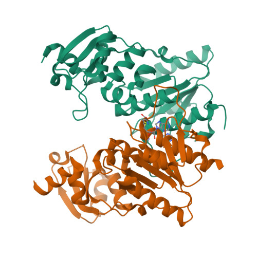
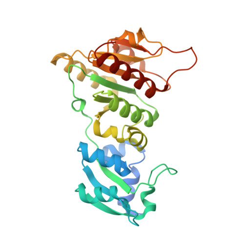
    The emergence of antibiotic-resistant bacterial strains is a widespread problem in medical practice and drug design, and each case requires the elucidation of the underlying mechanism. AviRb from Streptomyces viridochromogenes methylates the 2'-O atom of U2479 of the 23S ribosomal RNA in Gram-positive bacteria and thus mediates resistance to the oligosaccharide (orthosomycin) antibiotic avilamycin. The structure of AviRb with and without bound cofactor S-adenosyl-L-methionine (AdoMet) was determined, showing that it is a homodimer belonging to the SpoU family within the SPOUT class of methyltransferases. The relationships within this class were analyzed in detail and, in addition, a novel fourth SpoU sequence fingerprint is proposed. Each subunit of AviRb consists of two domains. The N-terminal domain, being related to the ribosomal proteins L30 and L7Ae, is likely to bind RNA. The C-terminal domain is related to all SPOUT methyltransferases, and is responsible for AdoMet-binding, catalysis and dimerization. The cofactor binds at the characteristic knot of the polypeptide in an unusually bent conformation. The transferred methyl group points to a broad cleft formed with the L30-type domain of the other subunit. Measurements of mutant activity revealed four important residues responsible for catalysis and allowed the modeling of a complex between AviRb and the RNA target. The model includes a specificity pocket for uracil but does not contain a base for deprotonating the 2'-O atom of U2479 on methylation.


  • Organizational Affiliation

    Institut für Organische Chemie und Biochemie, Albert-Ludwigs-Universität, Albertstr. 21, D-79104 Freiburg im Breisgau, Germany.


Macromolecules
Find similar proteins by:  (by identity cutoff)  |  3D Structure
Entity ID: 1
MoleculeChains Sequence LengthOrganismDetailsImage
rRNA methyltransferase
A, B
287Streptomyces viridochromogenesMutation(s): 0 
EC: 2.1.1 (PDB Primary Data), 2.1.1.208 (UniProt)
UniProt
Find proteins for Q9F5K6 (Streptomyces viridochromogenes)
Explore Q9F5K6 
Go to UniProtKB:  Q9F5K6
Entity Groups  
Sequence Clusters30% Identity50% Identity70% Identity90% Identity95% Identity100% Identity
UniProt GroupQ9F5K6
Sequence Annotations
Expand
  • Reference Sequence
Experimental Data & Validation

Experimental Data

  • Method: X-RAY DIFFRACTION
  • Resolution: 2.55 Å
  • R-Value Free:  0.257 (Depositor), 0.250 (DCC) 
  • R-Value Work:  0.202 (Depositor), 0.210 (DCC) 
  • R-Value Observed: 0.205 (Depositor) 
Space Group: P 41 21 2
Unit Cell:
Length ( Å )Angle ( ˚ )
a = 76.881α = 90
b = 76.881β = 90
c = 209.818γ = 90
Software Package:
Software NamePurpose
REFMACrefinement
XDSdata reduction
XDSdata scaling
AMoREphasing

Structure Validation

View Full Validation Report


Ligand Structure Quality Assessment 

Created with Raphaël 2.3.0Worse 01 BetterLigand structure goodness of fit to experimental dataBest fitted SAMClick on this verticalbar to view details

Entry History 

Deposition Data

Revision History  (Full details and data files)

  • Version 1.0: 2005-01-25
    Type: Initial release
  • Version 1.1: 2008-04-30
    Changes: Version format compliance
  • Version 1.2: 2011-07-13
    Changes: Advisory, Version format compliance
  • Version 1.3: 2014-04-16
    Changes: Experimental preparation
  • Version 1.4: 2017-10-11
    Changes: Refinement description
  • Version 1.5: 2024-03-13
    Changes: Data collection, Database references, Derived calculations