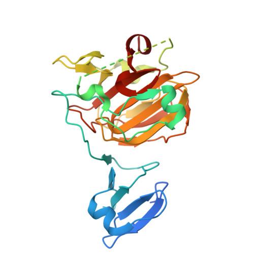
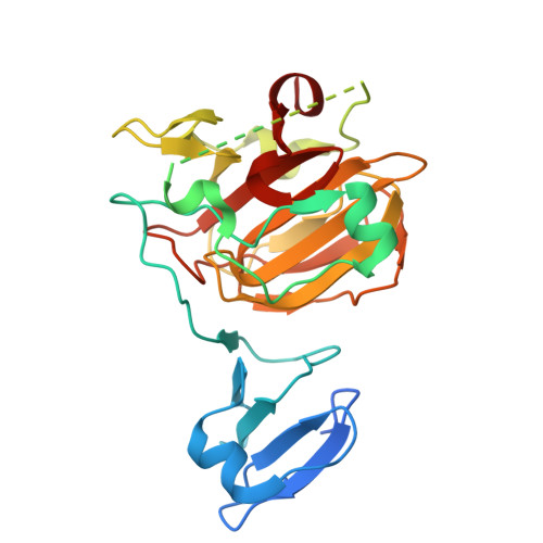
1QWY

Latent LytM at 1.3 A resolution


Experimental Data Snapshot

  • Method: X-RAY DIFFRACTION
  • Resolution: 1.30 Å
  • R-Value Free: 0.181 
  • R-Value Work: 0.163 
  • R-Value Observed: 0.164 

wwPDB Validation   3D Report Full Report


This is version 1.4 of the entry. See complete history


Literature

Latent LytM at 1.3A resolution.

Odintsov, S.G.Sabala, I.Marcyjaniak, M.Bochtler, M.

(2004) J Mol Biol 335: 775-785

  • DOI: https://doi.org/10.1016/j.jmb.2003.11.009
  • Primary Citation of Related Structures:  
    1QWY

  • PubMed Abstract: 

    LytM, an autolysin from Staphylococcus aureus, is a Zn(2+)-dependent glycyl-glycine endopeptidase with a characteristic HxH motif that belongs to the lysostaphin-type (MEROPS M23/37) of metallopeptidases. Here, we present the 1.3A crystal structure of LytM, the first structure of a lysostaphin-type peptidase. In the LytM structure, the Zn(2+) is tetrahedrally coordinated by the side-chains of N117, H210, D214 and H293, the second histidine of the HxH motif. Although close to the active-site, H291, the first histidine of the HxH motif, is not directly involved in Zn(2+)-coordination, and there is no water molecule in the coordination sphere of the Zn(2+), suggesting that the crystal structure shows a latent form of the enzyme. Although LytM has not previously been considered as a proenzyme, we show that a truncated version of LytM that lacks the N-terminal part with the poorly conserved Zn(2+) ligand N117 has much higher specific activity than full-length enzyme. This observation is consistent with the known removal of profragments in other lysostaphin-type proteins and with a prior observation of an active LytM degradation fragment in S.aureus supernatant. The "asparagine switch" in LytM is analogous to the "cysteine switch" in pro-matrix metalloproteases.


  • Organizational Affiliation

    International Institute of Molecular and Cell Biology, ul. Trojdena 4, 02-109 Warsaw, Poland.


Macromolecules
Find similar proteins by:  (by identity cutoff)  |  3D Structure
Entity ID: 1
MoleculeChains Sequence LengthOrganismDetailsImage
peptidoglycan hydrolase291Staphylococcus aureus subsp. aureus NCTC 8325Mutation(s): 0 
Gene Names: LytM
EC: 3.4.24.75
UniProt
Find proteins for O33599 (Staphylococcus aureus (strain NCTC 8325 / PS 47))
Explore O33599 
Go to UniProtKB:  O33599
Entity Groups  
Sequence Clusters30% Identity50% Identity70% Identity90% Identity95% Identity100% Identity
UniProt GroupO33599
Sequence Annotations
Expand
  • Reference Sequence
Small Molecules
Ligands 1 Unique
IDChains Name / Formula / InChI Key2D Diagram3D Interactions
ZN
Query on ZN

Download Ideal Coordinates CCD File 
B [auth A]ZINC ION
Zn
PTFCDOFLOPIGGS-UHFFFAOYSA-N
Experimental Data & Validation

Experimental Data

  • Method: X-RAY DIFFRACTION
  • Resolution: 1.30 Å
  • R-Value Free: 0.181 
  • R-Value Work: 0.163 
  • R-Value Observed: 0.164 
  • Space Group: P 1 21 1
Unit Cell:
Length ( Å )Angle ( ˚ )
a = 45.332α = 90
b = 53.232β = 104.02
c = 51.603γ = 90
Software Package:
Software NamePurpose
REFMACrefinement
MOSFLMdata reduction
CCP4data scaling
MLPHAREphasing

Structure Validation

View Full Validation Report


Entry History 

Deposition Data

Revision History  (Full details and data files)

  • Version 1.0: 2004-01-20
    Type: Initial release
  • Version 1.1: 2008-04-29
    Changes: Version format compliance
  • Version 1.2: 2011-07-13
    Changes: Version format compliance
  • Version 1.3: 2018-01-24
    Changes: Database references
  • Version 1.4: 2024-02-14
    Changes: Data collection, Database references, Derived calculations