Use of analogues of methionine and methionyl adenylate to sample conformational changes during catalysis in Escherichia coli methionyl-tRNA synthetase.
Crepin, T., Schmitt, E., Mechulam, Y., Sampson, P.B., Vaughan, M.D., Honek, J.F., Blanquet, S.(2003) J Mol Biol 332: 59-72
- PubMed: 12946347
- DOI: https://doi.org/10.1016/s0022-2836(03)00917-3
- Primary Citation of Related Structures:
1P7P, 1PFU, 1PFV, 1PFW, 1PFY, 1PG0, 1PG2 - PubMed Abstract:
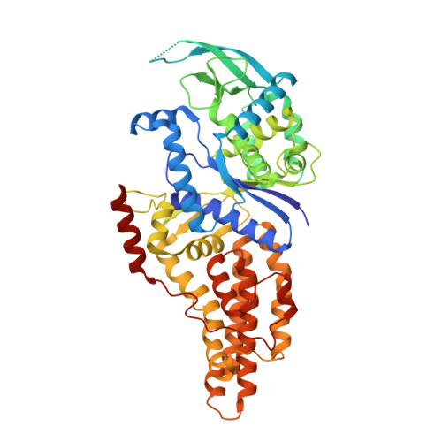
Binding of methionine to methionyl-tRNA synthetase (MetRS) is known to promote conformational changes within the active site. However, the contribution of these rearrangements to enzyme catalysis is not fully understood. In this study, several methionine and methionyl adenylate analogues were diffused into crystals of the monomeric form of Escherichia coli methionyl-tRNA synthetase. The structures of the corresponding complexes were solved at resolutions below 1.9A and compared to those of the enzyme free or complexed with methionine. Residues Y15 and W253 play key roles in the strength of the binding of the amino acid and of its analogues. Indeed, full motions of these residues are required to recover the maximum in free energy of binding. Residue Y15 also controls the size of the hydrophobic pocket where the amino acid side-chain interacts. H301 appears to participate to the specific recognition of the sulphur atom of methionine. Complexes with methionyl adenylate analogues illustrate the shielding by MetRS of the region joining the methionine and adenosine moieties. Finally, the structure of MetRS complexed to a methionine analogue mimicking the tetrahedral carbon of the transition state in the aminoacylation reaction was solved. On the basis of this model, we propose that, in response to the binding of the 3'-end of tRNA, Y15 moves again in order to deshield the anhydride bond in the natural adenylate.
Organizational Affiliation:
Laboratoire de Biochimie, Unité Mixte de Recherche no 7654, CNRS-Ecole Polytechnique, F-91128 Palaiseau cedex, France.


