1G87

THE CRYSTAL STRUCTURE OF ENDOGLUCANASE 9G FROM CLOSTRIDIUM CELLULOLYTICUM


Experimental Data Snapshot

  • Method: X-RAY DIFFRACTION
  • Resolution: 1.60 Å
  • R-Value Free: 0.200 
  • R-Value Work: 0.174 
  • R-Value Observed: 0.174 

wwPDB Validation   3D Report Full Report


This is version 1.4 of the entry. See complete history


Literature

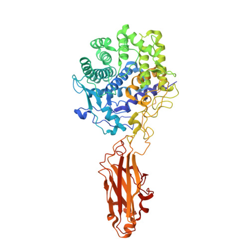
X-Ray Crystal Structure of the Multidomain Endoglucanase Cel9G from Clostridium cellulolyticum Complexed with Natural and Synthetic Cello-Oligosaccharides

Mandelman, D.Belaich, A.Belaich, J.P.Aghajari, N.Driguez, H.Haser, R.

(2003) J Bacteriol 185: 4127-4135

  • DOI: https://doi.org/10.1128/JB.185.14.4127-4135.2003
  • Primary Citation of Related Structures:  
    1G87, 1GA2, 1K72, 1KFG

  • PubMed Abstract: 

    Complete cellulose degradation is the first step in the use of biomass as a source of renewable energy. To this end, the engineering of novel cellulase activity, the activity responsible for the hydrolysis of the beta-1,4-glycosidic bonds in cellulose, is a topic of great interest. The high-resolution X-ray crystal structure of a multidomain endoglucanase from Clostridium cellulolyticum has been determined at a 1.6-A resolution. The endoglucanase, Cel9G, is comprised of a family 9 catalytic domain attached to a family III(c) cellulose-binding domain. The two domains together form a flat platform onto which crystalline cellulose is suggested to bind and be fed into the active-site cleft for endolytic hydrolysis. To further dissect the structural basis of cellulose binding and hydrolysis, the structures of Cel9G in the presence of cellobiose, cellotriose, and a DP-10 thio-oligosaccharide inhibitor were resolved at resolutions of 1.7, 1.8, and 1.9 A, respectively.


  • Organizational Affiliation

    Laboratoire de BioCristallographie, Institut de Biologie et Chimie des Protéines, UMR 5086, CNRS et Université Claude Bernard Lyon I, 69367 Lyon, Cedex 07, France.


Macromolecules
Find similar proteins by:  (by identity cutoff)  |  3D Structure
Entity ID: 1
MoleculeChains Sequence LengthOrganismDetailsImage
ENDOCELLULASE 9G
A, B
614Ruminiclostridium cellulolyticumMutation(s): 0 
Gene Names: CELCCG
EC: 3.2.1.4
UniProt
Find proteins for P37700 (Ruminiclostridium cellulolyticum (strain ATCC 35319 / DSM 5812 / JCM 6584 / H10))
Explore P37700 
Go to UniProtKB:  P37700
Entity Groups  
Sequence Clusters30% Identity50% Identity70% Identity90% Identity95% Identity100% Identity
UniProt GroupP37700
Sequence Annotations
Expand
  • Reference Sequence
Small Molecules
Ligands 4 Unique
IDChains Name / Formula / InChI Key2D Diagram3D Interactions
GOL
Query on GOL

Download Ideal Coordinates CCD File 
CA [auth B]
DA [auth B]
K [auth A]
L [auth A]
M [auth A]
CA [auth B],
DA [auth B],
K [auth A],
L [auth A],
M [auth A],
N [auth A]
GLYCEROL
C3 H8 O3
PEDCQBHIVMGVHV-UHFFFAOYSA-N
EDO
Query on EDO

Download Ideal Coordinates CCD File 
AA [auth B]
BA [auth B]
H [auth A]
I [auth A]
J [auth A]
AA [auth B],
BA [auth B],
H [auth A],
I [auth A],
J [auth A],
U [auth B],
V [auth B],
W [auth B],
X [auth B],
Y [auth B],
Z [auth B]
1,2-ETHANEDIOL
C2 H6 O2
LYCAIKOWRPUZTN-UHFFFAOYSA-N
CA
Query on CA

Download Ideal Coordinates CCD File 
C [auth A],
D [auth A],
O [auth B],
P [auth B]
CALCIUM ION
Ca
BHPQYMZQTOCNFJ-UHFFFAOYSA-N
MG
Query on MG

Download Ideal Coordinates CCD File 
E [auth A]
F [auth A]
G [auth A]
Q [auth B]
R [auth B]
E [auth A],
F [auth A],
G [auth A],
Q [auth B],
R [auth B],
S [auth B],
T [auth B]
MAGNESIUM ION
Mg
JLVVSXFLKOJNIY-UHFFFAOYSA-N
Experimental Data & Validation

Experimental Data

  • Method: X-RAY DIFFRACTION
  • Resolution: 1.60 Å
  • R-Value Free: 0.200 
  • R-Value Work: 0.174 
  • R-Value Observed: 0.174 
  • Space Group: P 1
Unit Cell:
Length ( Å )Angle ( ˚ )
a = 56.85α = 94.22
b = 57.72β = 100.87
c = 86.27γ = 99.61
Software Package:
Software NamePurpose
MAR345data collection
SCALEPACKdata scaling
AMoREphasing
ARP/wARPmodel building
CNSrefinement

Structure Validation

View Full Validation Report


Entry History 

Deposition Data

Revision History  (Full details and data files)

  • Version 1.0: 2003-07-15
    Type: Initial release
  • Version 1.1: 2008-04-01
    Changes: Version format compliance
  • Version 1.2: 2011-07-13
    Changes: Non-polymer description, Version format compliance
  • Version 1.3: 2017-10-04
    Changes: Refinement description
  • Version 1.4: 2023-08-09
    Changes: Data collection, Database references, Derived calculations, Refinement description