1E3P

tungstate derivative of Streptomyces antibioticus PNPase/GPSI enzyme


Experimental Data Snapshot

  • Method: X-RAY DIFFRACTION
  • Resolution: 2.50 Å
  • R-Value Free: 0.247 
  • R-Value Work: 0.213 
  • R-Value Observed: 0.213 

wwPDB Validation   3D Report Full Report


This is version 1.4 of the entry. See complete history


Literature

A Duplicated Fold is the Structural Basis for Polynucleotide Phosphorylase Catalytic Activity, Processivity, and Regulation

Symmons, M.F.Jones, G.H.Luisi, B.F.

(2000) Structure 8: 1215

  • DOI: https://doi.org/10.1016/s0969-2126(00)00521-9
  • Primary Citation of Related Structures:  
    1E3H, 1E3P

  • PubMed Abstract: 

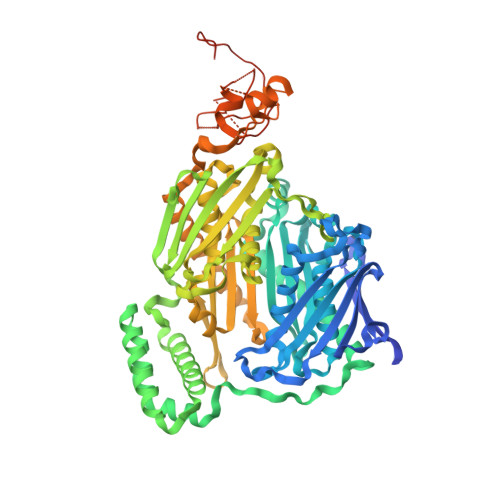
    Polynucleotide phosphorylase (PNPase) is a polyribonucleotide nucleotidyl transferase (E.C.2.7.7.8) that degrades mRNA in prokaryotes. Streptomyces antibioticus PNPase also assays as a guanosine 3'-diphosphate 5'-triphosphate (pppGpp) synthetase (E.C.2.7.6.5). It may function to coordinate changes in mRNA lifetimes with pppGpp levels during the Streptomyces lifecycle. The structure of S. antibioticus PNPase without bound RNA but with the phosphate analog tungstate bound at the PNPase catalytic sites was determined by X-ray crystallography and shows a trimeric multidomain protein with a central channel. The structural core has a novel duplicated architecture formed by association of two homologous domains. The tungstate derivative structure reveals the PNPase active site in the second of these core domains. Structure-based sequence analysis suggests that the pppGpp synthetase active site is located in the first core domain. This is the first structure of a PNPase and shows the structural basis for the trimer assembly, the arrangement of accessory RNA binding domains, and the likely catalytic residues of the PNPase active site. A possible function of the trimer channel is as a contribution to both the processivity of degradation and the regulation of PNPase action by RNA structural elements.


  • Organizational Affiliation

    Department of Biochemistry, University of Cambridge, Cambridge, United Kingdom. mfs@mole.bio.cam.ac.uk


Macromolecules
Find similar proteins by:  (by identity cutoff)  |  3D Structure
Entity ID: 1
MoleculeChains Sequence LengthOrganismDetailsImage
Polyribonucleotide nucleotidyltransferase757Streptomyces antibioticusMutation(s): 0 
Gene Names: pnpAFM16_28085
EC: 2.7.7.8
UniProt
Find proteins for Q53597 (Streptomyces antibioticus)
Explore Q53597 
Go to UniProtKB:  Q53597
Entity Groups  
Sequence Clusters30% Identity50% Identity70% Identity90% Identity95% Identity100% Identity
UniProt GroupQ53597
Sequence Annotations
Expand
  • Reference Sequence
Small Molecules
Ligands 2 Unique
IDChains Name / Formula / InChI Key2D Diagram3D Interactions
WO4
Query on WO4

Download Ideal Coordinates CCD File 
L [auth A]TUNGSTATE(VI)ION
O4 W
PBYZMCDFOULPGH-UHFFFAOYSA-N
SO4
Query on SO4

Download Ideal Coordinates CCD File 
B [auth A]
C [auth A]
D [auth A]
E [auth A]
F [auth A]
B [auth A],
C [auth A],
D [auth A],
E [auth A],
F [auth A],
G [auth A],
H [auth A],
I [auth A],
J [auth A],
K [auth A]
SULFATE ION
O4 S
QAOWNCQODCNURD-UHFFFAOYSA-L
Experimental Data & Validation

Experimental Data

  • Method: X-RAY DIFFRACTION
  • Resolution: 2.50 Å
  • R-Value Free: 0.247 
  • R-Value Work: 0.213 
  • R-Value Observed: 0.213 
  • Space Group: H 3 2
Unit Cell:
Length ( Å )Angle ( ˚ )
a = 130.83α = 90
b = 130.83β = 90
c = 328.732γ = 120
Software Package:
Software NamePurpose
CNSrefinement
DENZOdata reduction
SCALEPACKdata scaling
CNSphasing

Structure Validation

View Full Validation Report


Entry History 

Deposition Data

Revision History  (Full details and data files)

  • Version 1.0: 2000-11-03
    Type: Initial release
  • Version 1.1: 2011-05-08
    Changes: Version format compliance
  • Version 1.2: 2011-07-13
    Changes: Version format compliance
  • Version 1.3: 2019-11-06
    Changes: Advisory, Data collection, Database references, Other, Source and taxonomy, Structure summary
  • Version 1.4: 2023-12-13
    Changes: Data collection, Database references, Derived calculations, Refinement description