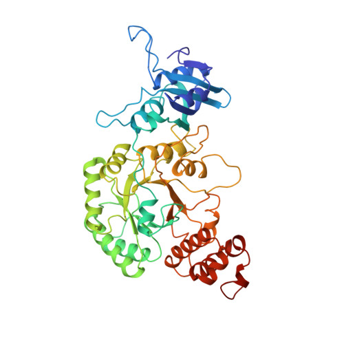
Crystal structure of activated ribulose-1,5-bisphosphate carboxylase complexed with its substrate, ribulose-1,5-bisphosphate.
Lundqvist, T., Schneider, G.(1991) J Biol Chem 266: 12604-12611
- PubMed: 1905726
- Primary Citation of Related Structures:
9RUB - PubMed Abstract:
The three-dimensional structure of the complex of ribulose-1,5-bisphosphate carboxylase from Rhodospirillum rubrum, CO2, Mg2+, and ribulose bisphosphate has been determined with x-ray crystallographic methods to 2.6-A resolution. Ribulose-1,5-bisphosphate binds across the active site with the two phosphate groups in the two phosphate binding sites of the beta/alpha barrel. The oxygen atoms of the carbamate and the side chain of Asp-193 provide the protein ligands to the bound Mg2+ ion. The C2 and the C3 or C4 oxygen atoms of the substrate are also within the first coordination sphere of the metal ion. At the present resolution of the electron density maps, two slightly different conformations of the substrate, with the C3 hydroxyl group "cis" or "trans" to the C2 oxygen, can be built into the observed electron density. The two different conformations suggest two different mechanisms of proton abstraction in the first step of catalysis, the enolization of the ribulose 1,5-bisphosphate. Two loop regions, which are disordered in the crystals of the nonactivated enzyme, could be built into their respective electron density. A comparison with the structure of the quaternary complex of the spinach enzyme shows that despite the different conformations of loop 6, the positions of the Mg2+ ion, and most atoms of the substrate are very similar when superimposed on each other. There are, however, some significant differences at the active site, especially in the metal coordination sphere.
Organizational Affiliation:
Department of Molecular Biology, Swedish University of Agricultural Sciences, Uppsala.