RCSB PDB - 9JF5: Arginine decarboxylase in Aspergillus oryzae complexed with arginine

 9JF5

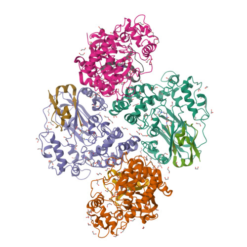
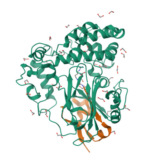
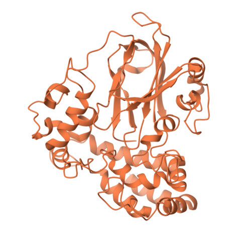
Arginine decarboxylase in Aspergillus oryzae complexed with arginine


Experimental Data Snapshot

  • Method: X-RAY DIFFRACTION
  • Resolution: 1.90 Å
  • R-Value Free: 0.189 
  • R-Value Work: 0.160 
  • R-Value Observed: 0.162 

Starting Model: in silico
View more details

wwPDB Validation   3D Report Full Report


Ligand Structure Quality Assessment 

Created with Raphaël 2.3.0Worse 01 BetterLigand structure goodness of fit to experimental dataBest fitted AG2Click on this verticalbar to view details

This is version 1.0 of the entry. See complete history


Literature

Unveiling the reaction mechanism of arginine decarboxylase in Aspergillus oryzae: Insights from crystal structure analysis.

Odagaki, Y.Murakami, Y.Takita, T.Mizutani, K.Mikami, B.Fujiwara, S.Yasukawa, K.

(2024) Biochem Biophys Res Commun 733: 150728-150728

  • DOI: https://doi.org/10.1016/j.bbrc.2024.150728
  • Primary Citation of Related Structures:  
    9JER, 9JF5, 9JFN

  • PubMed Abstract: 

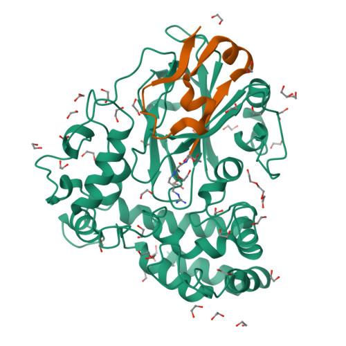
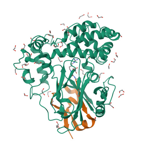
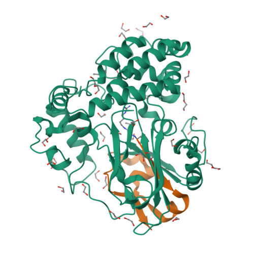
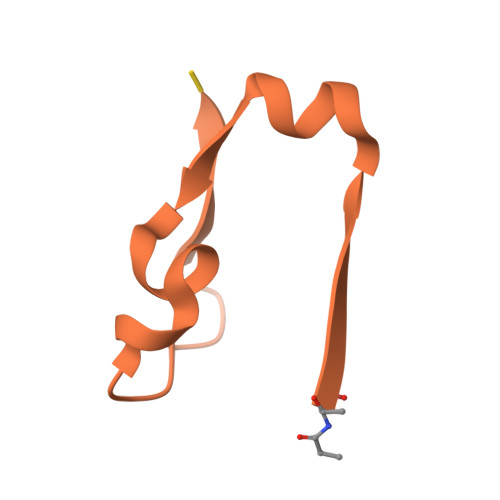
    Agmatine, a natural polyamine also known as 4-aminobutyl-guanidine, is biosynthesized from arginine by decarboxylation. Aspergillus oryzae contains high amounts of agmatine, suggesting highly active arginine decarboxylase (ADC) in this organism. However, genome analysis revealed no ADC homolog in A. oryzae. A. oryzae strain RIB40 has six homologs of phosphatidylserine decarboxylase (PSD), an enzyme that synthesizes phosphatidyl ethanolamine from phosphatidylserine. We previously discovered that one of these homologs, AO090102000327, encodes arginine decarboxylase, which we named ADC1. In the present study, we determined the crystal structures of ligand-free, arginine-treated, and agmatine-treated ADC1 each at 1.9-2.15 Å resolution. Each structure contained four ADC1 molecules (chains A-D) in the asymmetric unit of the cell. Each ADC1 molecule is a heterodimer consisting of the N-terminal region (Asn60-Gly441) and C-terminal region (Ser442-Thr482). In the ligand-free ADC1, the N-terminus of Ser442 was modified to form a pyruvoyl group. In the arginine-treated ADC1, arginine was converted to agmatine, with the pyruvoyl group covalently bound to agmatine by forming a Schiff base. The same structure was observed in agmatine-treated ADC1. These results indicate that ADC1 is a pyruvoyl-dependent decarboxylase and unveils the reaction mechanism of ADC from A. oryzae.


  • Organizational Affiliation

    Division of Food Science and Biotechnology, Graduate School of Agriculture, Kyoto University, Sakyo-ku, Kyoto, 606-8502, Japan.


Macromolecules
Find similar proteins by:  (by identity cutoff)  |  3D Structure
Entity ID: 1
MoleculeChains Sequence LengthOrganismDetailsImage
L-tryptophan decarboxylase PsiD-like domain-containing proteinA,
C [auth B],
E [auth C],
G [auth D]
462Aspergillus oryzae RIB40Mutation(s): 0 
Gene Names: AO090102000327
EC: 4.1.1.19
UniProt
Find proteins for Q2UAM5 (Aspergillus oryzae (strain ATCC 42149 / RIB 40))
Explore Q2UAM5 
Go to UniProtKB:  Q2UAM5
Entity Groups  
Sequence Clusters30% Identity50% Identity70% Identity90% Identity95% Identity100% Identity
UniProt GroupQ2UAM5
Sequence Annotations
Expand
  • Reference Sequence
Find similar proteins by:  (by identity cutoff)  |  3D Structure
Entity ID: 2
MoleculeChains Sequence LengthOrganismDetailsImage
L-tryptophan decarboxylase PsiD-like domain-containing proteinB [auth E],
D [auth F],
F [auth G],
H
42Aspergillus oryzae RIB40Mutation(s): 1 
Gene Names: AO090102000327
EC: 4.1.1.19
UniProt
Find proteins for Q2UAM5 (Aspergillus oryzae (strain ATCC 42149 / RIB 40))
Explore Q2UAM5 
Go to UniProtKB:  Q2UAM5
Entity Groups  
Sequence Clusters30% Identity50% Identity70% Identity90% Identity95% Identity100% Identity
UniProt GroupQ2UAM5
Sequence Annotations
Expand
  • Reference Sequence
Small Molecules
Ligands 3 Unique
IDChains Name / Formula / InChI Key2D Diagram3D Interactions
AG2 (Subject of Investigation/LOI)
Query on AG2

Download Ideal Coordinates CCD File 
KF [auth H],
LC [auth F],
SA [auth E],
YD [auth G]
AGMATINE
C5 H14 N4
QYPPJABKJHAVHS-UHFFFAOYSA-N
PEG
Query on PEG

Download Ideal Coordinates CCD File 
BE [auth D]
I [auth A]
J [auth A]
NC [auth C]
OC [auth C]
DI(HYDROXYETHYL)ETHER
C4 H10 O3
MTHSVFCYNBDYFN-UHFFFAOYSA-N
EDO
Query on EDO

Download Ideal Coordinates CCD File 
AA [auth A]
AB [auth B]
AC [auth B]
AD [auth C]
AE [auth G]
1,2-ETHANEDIOL
C2 H6 O2
LYCAIKOWRPUZTN-UHFFFAOYSA-N
Experimental Data & Validation

Experimental Data

  • Method: X-RAY DIFFRACTION
  • Resolution: 1.90 Å
  • R-Value Free: 0.189 
  • R-Value Work: 0.160 
  • R-Value Observed: 0.162 
  • Space Group: C 2 2 21
Unit Cell:
Length ( Å )Angle ( ˚ )
a = 134.02α = 90
b = 195.575β = 90
c = 196.124γ = 90
Software Package:
Software NamePurpose
PHENIXrefinement
XDSdata reduction
XDSdata scaling
PHENIXphasing

Structure Validation

View Full Validation Report


Ligand Structure Quality Assessment 

Created with Raphaël 2.3.0Worse 01 BetterLigand structure goodness of fit to experimental dataBest fitted AG2Click on this verticalbar to view details

Entry History & Funding Information

Deposition Data


Funding OrganizationLocationGrant Number
Japan Science and TechnologyJapan23K24590
Japan Science and TechnologyJapan22H03332
Japan Science and TechnologyJapan22K05441

Revision History  (Full details and data files)

  • Version 1.0: 2024-10-09
    Type: Initial release