8ZES

Crystal structure of the Wuhan SARS-CoV-2 RBD (333-541) complexed with P2C5 nanobody


Experimental Data Snapshot

  • Method: X-RAY DIFFRACTION
  • Resolution: 3.70 Å
  • R-Value Free: 0.269 
  • R-Value Work: 0.207 
  • R-Value Observed: 0.211 

Starting Models: experimental, in silico
View more details

wwPDB Validation   3D Report Full Report


Ligand Structure Quality Assessment 


This is version 1.2 of the entry. See complete history


Literature

Structural Basis for Evasion of New SARS-CoV-2 Variants from the Potent Virus-Neutralizing Nanobody Targeting the S-Protein Receptor-Binding Domain.

Sluchanko, N.N.Shcheblyakov, D.V.Varfolomeeva, L.A.Favorskaya, I.A.Dolzhikova, I.V.Korobkova, A.I.Alekseeva, I.A.Esmagambetov, I.B.Derkaev, A.A.Prokofiev, V.V.Zorkov, I.D.Logunov, D.Y.Gintsburg, A.L.Popov, V.O.Boyko, K.M.

(2024) Biochemistry (Mosc) 89: 1260-1272

  • DOI: https://doi.org/10.1134/S0006297924070083
  • Primary Citation of Related Structures:  
    8ZER, 8ZES

  • PubMed Abstract: 

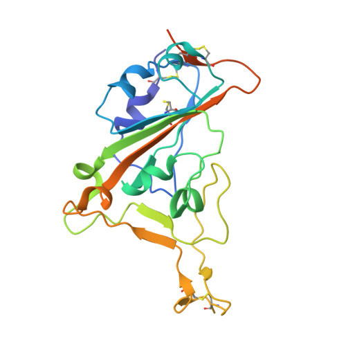
    COVID-19 has caused millions of deaths and many times more infections worldwide, emphasizing the unpreparedness of the global health system in the face of new infections and the key role for vaccines and therapeutics, including virus-neutralizing antibodies, in prevention and containment of the disease. Continuous evolution of the SARS-CoV-2 coronavirus has been causing its new variants to evade the action of the immune system, which highlighted the importance of detailed knowledge of the epitopes of already selected potent virus-neutralizing antibodies. A single-chain antibody ("nanobody") targeting the SARS-CoV-2 receptor-binding domain (RBD), clone P2C5, had exhibited robust virus-neutralizing activity against all SARS-CoV-2 variants and, being a major component of the anti-COVID-19 formulation "GamCoviMab", had successfully passed Phase I of clinical trials. However, after the emergence of the Delta and XBB variants, a decrease in the neutralizing activity of this nanobody was observed. Here we report on the successful crystal structure determination of the RBD:P2C5 complex at 3.1 Å, which revealed the intricate protein-protein interface, sterically occluding full ACE2 receptor binding by the P2C5-neutralized RBD. Moreover, the structure revealed the developed RBD:P2C5 interface centered around residues Leu452 and Phe490, thereby explaining the evasion of the Delta or Omicron XBB, but not Omicron B.1.1.529 variant, as a result of the single L452R or F490S mutations, respectively, from the action of P2C5. The structure obtained is expected to foster nanobody engineering in order to rescue neutralization activity and will facilitate epitope mapping for other neutralizing nanobodies by competition assays.


  • Organizational Affiliation

    Bach Institute of Biochemistry, Federal Research Centre "Fundamentals of Biotechnology", Russian Academy of Sciences, Moscow, 119071, Russia. nikolai.sluchanko@mail.ru.


Macromolecules
Find similar proteins by:  (by identity cutoff)  |  3D Structure
Entity ID: 1
MoleculeChains Sequence LengthOrganismDetailsImage
Spike protein S1A [auth C],
C [auth A],
F [auth G],
H [auth I],
J [auth D]
221Severe acute respiratory syndrome coronavirus 2Mutation(s): 0 
Gene Names: S2
UniProt
Find proteins for P0DTC2 (Severe acute respiratory syndrome coronavirus 2)
Explore P0DTC2 
Go to UniProtKB:  P0DTC2
Entity Groups  
Sequence Clusters30% Identity50% Identity70% Identity90% Identity95% Identity100% Identity
UniProt GroupP0DTC2
Glycosylation
Glycosylation Sites: 1Go to GlyGen: P0DTC2-1
Sequence Annotations
Expand
  • Reference Sequence
Find similar proteins by:  (by identity cutoff)  |  3D Structure
Entity ID: 2
MoleculeChains Sequence LengthOrganismDetailsImage
Nanobody P2C5B [auth E],
D [auth B],
E [auth F],
G [auth H],
I [auth J]
146Camelus bactrianusMutation(s): 0 
Entity Groups  
Sequence Clusters30% Identity50% Identity70% Identity90% Identity95% Identity100% Identity
Sequence Annotations
Expand
  • Reference Sequence
Oligosaccharides

Help

Entity ID: 3
MoleculeChains Length2D Diagram Glycosylation3D Interactions
2-acetamido-2-deoxy-beta-D-glucopyranose-(1-4)-2-acetamido-2-deoxy-beta-D-glucopyranose
K, L, M
2N-Glycosylation
Glycosylation Resources
GlyTouCan:  G42666HT
GlyCosmos:  G42666HT
GlyGen:  G42666HT
Experimental Data & Validation

Experimental Data

  • Method: X-RAY DIFFRACTION
  • Resolution: 3.70 Å
  • R-Value Free: 0.269 
  • R-Value Work: 0.207 
  • R-Value Observed: 0.211 
  • Space Group: I 21 21 21
Unit Cell:
Length ( Å )Angle ( ˚ )
a = 126.64α = 90
b = 194.24β = 90
c = 264.37γ = 90
Software Package:
Software NamePurpose
REFMACrefinement
XDSdata reduction
XSCALEdata scaling
MOLREPphasing

Structure Validation

View Full Validation Report


Ligand Structure Quality Assessment 


Entry History & Funding Information

Deposition Data


Funding OrganizationLocationGrant Number
Not funded--

Revision History  (Full details and data files)

  • Version 1.0: 2024-09-04
    Type: Initial release
  • Version 1.1: 2024-09-18
    Changes: Database references
  • Version 1.2: 2024-10-16
    Changes: Structure summary