8Y3X

Cell divisome sPG hydrolysis machinery FtsEX-EnvC


Experimental Data Snapshot

  • Method: ELECTRON MICROSCOPY
  • Resolution: 3.11 Å
  • Aggregation State: PARTICLE 
  • Reconstruction Method: SINGLE PARTICLE 

wwPDB Validation   3D Report Full Report


This is version 1.1 of the entry. See complete history


Literature

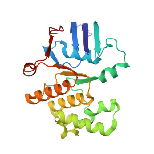
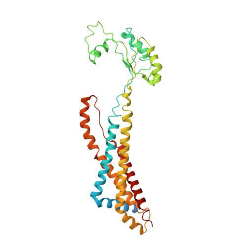
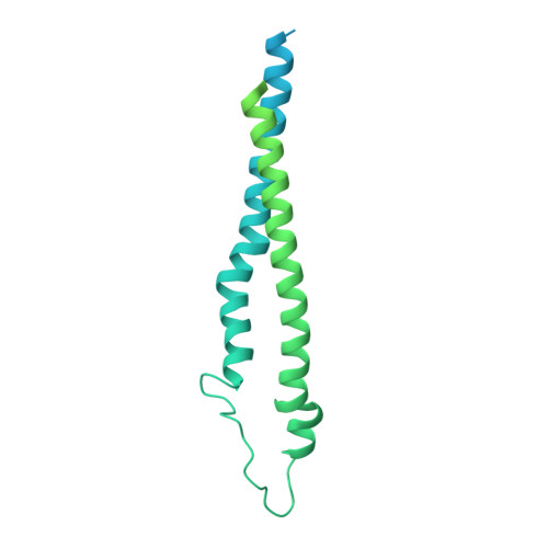
Structure and activity of the septal peptidoglycan hydrolysis machinery crucial for bacterial cell division.

Chen, Y.Gu, J.Yang, B.Yang, L.Pang, J.Luo, Q.Li, Y.Li, D.Deng, Z.Dong, C.Dong, H.Zhang, Z.

(2024) PLoS Biol 22: e3002628-e3002628

  • DOI: https://doi.org/10.1371/journal.pbio.3002628
  • Primary Citation of Related Structures:  
    8X61, 8Y3X

  • PubMed Abstract: 

    The peptidoglycan (PG) layer is a critical component of the bacterial cell wall and serves as an important target for antibiotics in both gram-negative and gram-positive bacteria. The hydrolysis of septal PG (sPG) is a crucial step of bacterial cell division, facilitated by FtsEX through an amidase activation system. In this study, we present the cryo-EM structures of Escherichia coli FtsEX and FtsEX-EnvC in the ATP-bound state at resolutions of 3.05 Å and 3.11 Å, respectively. Our PG degradation assays in E. coli reveal that the ATP-bound conformation of FtsEX activates sPG hydrolysis of EnvC-AmiB, whereas EnvC-AmiB alone exhibits autoinhibition. Structural analyses indicate that ATP binding induces conformational changes in FtsEX-EnvC, leading to significant differences from the apo state. Furthermore, PG degradation assays of AmiB mutants confirm that the regulation of AmiB by FtsEX-EnvC is achieved through the interaction between EnvC-AmiB. These findings not only provide structural insight into the mechanism of sPG hydrolysis and bacterial cell division, but also have implications for the development of novel therapeutics targeting drug-resistant bacteria.


  • Organizational Affiliation

    Department of Clinical Laboratory, Zhongnan Hospital of Wuhan University, School of Pharmaceutical Sciences, Wuhan University, Wuhan, China.


Macromolecules
Find similar proteins by:  (by identity cutoff)  |  3D Structure
Entity ID: 1
MoleculeChains Sequence LengthOrganismDetailsImage
Cell division ATP-binding protein FtsE
A, B
222Escherichia coliMutation(s): 1 
Gene Names: ftsEb3463JW3428
Membrane Entity: Yes 
UniProt
Find proteins for P0A9R7 (Escherichia coli (strain K12))
Explore P0A9R7 
Go to UniProtKB:  P0A9R7
Entity Groups  
Sequence Clusters30% Identity50% Identity70% Identity90% Identity95% Identity100% Identity
UniProt GroupP0A9R7
Sequence Annotations
Expand
  • Reference Sequence
Find similar proteins by:  (by identity cutoff)  |  3D Structure
Entity ID: 2
MoleculeChains Sequence LengthOrganismDetailsImage
Cell division protein FtsX
C, D
352Escherichia coliMutation(s): 0 
Gene Names: ftsXftsSb3462JW3427
Membrane Entity: Yes 
UniProt
Find proteins for P0AC30 (Escherichia coli (strain K12))
Explore P0AC30 
Go to UniProtKB:  P0AC30
Entity Groups  
Sequence Clusters30% Identity50% Identity70% Identity90% Identity95% Identity100% Identity
UniProt GroupP0AC30
Sequence Annotations
Expand
  • Reference Sequence
Find similar proteins by:  (by identity cutoff)  |  3D Structure
Entity ID: 3
MoleculeChains Sequence LengthOrganismDetailsImage
Murein hydrolase activator EnvC419Escherichia coliMutation(s): 0 
Gene Names: envCyibPb3613JW5646
Membrane Entity: Yes 
UniProt
Find proteins for P37690 (Escherichia coli (strain K12))
Explore P37690 
Go to UniProtKB:  P37690
Entity Groups  
Sequence Clusters30% Identity50% Identity70% Identity90% Identity95% Identity100% Identity
UniProt GroupP37690
Sequence Annotations
Expand
  • Reference Sequence
Experimental Data & Validation

Experimental Data

  • Method: ELECTRON MICROSCOPY
  • Resolution: 3.11 Å
  • Aggregation State: PARTICLE 
  • Reconstruction Method: SINGLE PARTICLE 

Structure Validation

View Full Validation Report


Entry History & Funding Information

Deposition Data


Funding OrganizationLocationGrant Number
National Natural Science Foundation of China (NSFC)China31800052

Revision History  (Full details and data files)

  • Version 1.0: 2024-05-08
    Type: Initial release
  • Version 1.1: 2024-06-19
    Changes: Database references