8U6S

Crystal Structure of HIV-1 Reverse Transcriptase in Complex with 8-(2-(3-morpholino-3-oxopropoxy)phenoxy)indolizine-2-carbonitrile (JLJ757), a non-nucleoside inhibitor


Experimental Data Snapshot

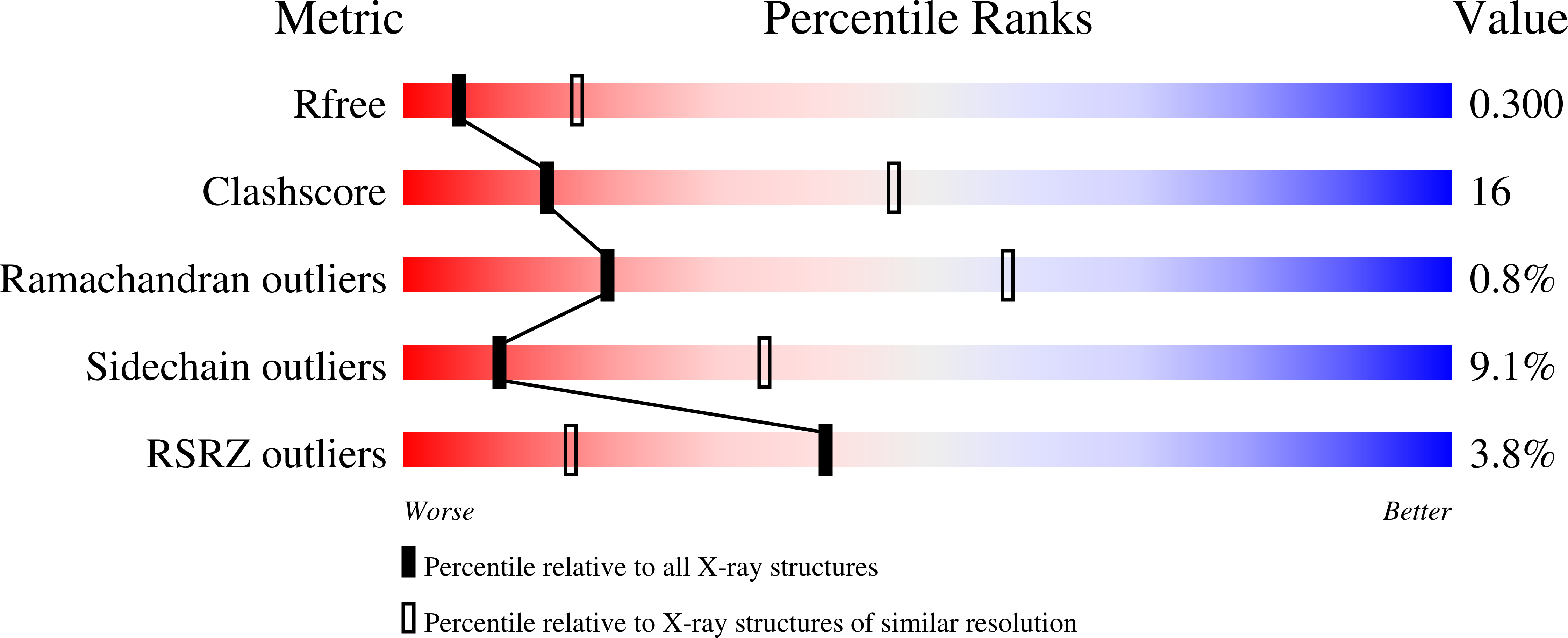
  • Method: X-RAY DIFFRACTION
  • Resolution: 2.99 Å
  • R-Value Free: 0.299 
  • R-Value Work: 0.225 
  • R-Value Observed: 0.229 

Starting Model: experimental
View more details

wwPDB Validation   3D Report Full Report


Ligand Structure Quality Assessment 


This is version 1.0 of the entry. See complete history


Literature

Covalent and noncovalent strategies for targeting Lys102 in HIV-1 reverse transcriptase.

Prucha, G.R.Henry, S.Hollander, K.Carter, Z.J.Spasov, K.A.Jorgensen, W.L.Anderson, K.S.

(2023) Eur J Med Chem 262: 115894-115894

  • DOI: https://doi.org/10.1016/j.ejmech.2023.115894
  • Primary Citation of Related Structures:  
    8U69, 8U6A, 8U6B, 8U6C, 8U6D, 8U6E, 8U6F, 8U6G, 8U6H, 8U6I, 8U6J, 8U6K, 8U6L, 8U6M, 8U6N, 8U6O, 8U6P, 8U6Q, 8U6R, 8U6S, 8U6T

  • PubMed Abstract: 

    Reverse transcriptase (RT) is one of three key proteins responsible for the replication cycle of HIV-1 in the host. Several classes of inhibitors have been developed to target the enzyme, with non-nucleoside reverse transcriptase inhibitors forming first-line treatment. Previously, covalent RT inhibitors have been identified and found to bind irreversibly to commonly mutated residues such as Y181C. In this work we aim to circumvent the issue of NNRTI resistance through targeting K102, which has not yet been identified to confer drug resistance. As reported here, 34 compounds were synthesized and characterized biochemically and structurally with wild-type (WT) HIV-1 RT. Two of these inhibitors demonstrate covalent inhibition as evidenced by protein crystallography, enzyme kinetics, mass spectrometry, and antiviral potency in HIV-1 infected human T-cell assays.


  • Organizational Affiliation

    Department of Pharmacology, Yale University School of Medicine, New Haven, CT, 06520-8066, USA.


Macromolecules
Find similar proteins by:  (by identity cutoff)  |  3D Structure
Entity ID: 1
MoleculeChains Sequence LengthOrganismDetailsImage
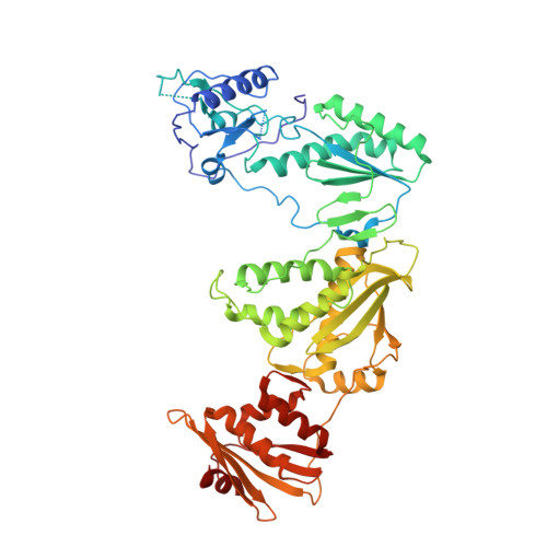
Reverse transcriptase/ribonuclease H554Human immunodeficiency virus 1Mutation(s): 3 
Gene Names: gag-pol
EC: 2.7.7.49 (PDB Primary Data), 2.7.7.7 (PDB Primary Data), 3.1.26.13 (PDB Primary Data)
UniProt
Find proteins for P03366 (Human immunodeficiency virus type 1 group M subtype B (isolate BH10))
Explore P03366 
Go to UniProtKB:  P03366
Entity Groups  
Sequence Clusters30% Identity50% Identity70% Identity90% Identity95% Identity100% Identity
UniProt GroupP03366
Sequence Annotations
Expand
  • Reference Sequence
Find similar proteins by:  (by identity cutoff)  |  3D Structure
Entity ID: 2
MoleculeChains Sequence LengthOrganismDetailsImage
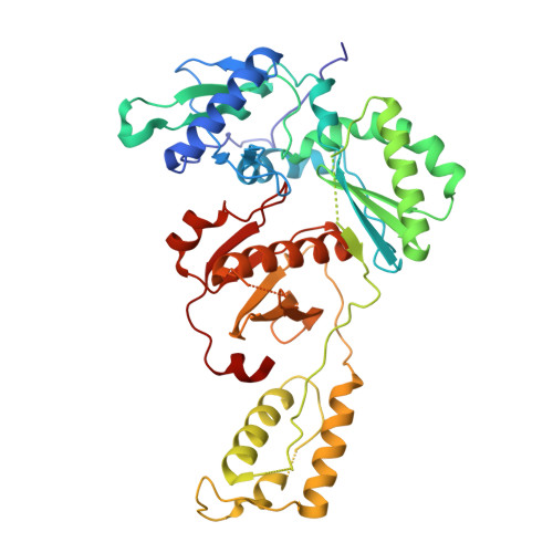
p51 RT428Human immunodeficiency virus 1Mutation(s): 1 
Gene Names: gag-pol
UniProt
Find proteins for P03366 (Human immunodeficiency virus type 1 group M subtype B (isolate BH10))
Explore P03366 
Go to UniProtKB:  P03366
Entity Groups  
Sequence Clusters30% Identity50% Identity70% Identity90% Identity95% Identity100% Identity
UniProt GroupP03366
Sequence Annotations
Expand
  • Reference Sequence
Experimental Data & Validation

Experimental Data

  • Method: X-RAY DIFFRACTION
  • Resolution: 2.99 Å
  • R-Value Free: 0.299 
  • R-Value Work: 0.225 
  • R-Value Observed: 0.229 
  • Space Group: C 1 2 1
Unit Cell:
Length ( Å )Angle ( ˚ )
a = 222.126α = 90
b = 68.624β = 107.41
c = 103.701γ = 90
Software Package:
Software NamePurpose
PHENIXrefinement
XDSdata reduction
Aimlessdata scaling
PHASERphasing

Structure Validation

View Full Validation Report


Ligand Structure Quality Assessment 


Entry History & Funding Information

Deposition Data


Funding OrganizationLocationGrant Number
National Institutes of Health/National Institute of General Medical Sciences (NIH/NIGMS)United StatesNIH/NAD AI155072

Revision History  (Full details and data files)

  • Version 1.0: 2023-11-08
    Type: Initial release