8RMO

Crystal structure of anti-FLAG M2 Fab fragment bound to FLAG-tag peptide epitope


Experimental Data Snapshot

  • Method: X-RAY DIFFRACTION
  • Resolution: 1.16 Å
  • R-Value Free: 0.185 
  • R-Value Work: 0.158 

wwPDB Validation   3D Report Full Report


This is version 1.1 of the entry. See complete history


Literature

Structural basis for recognition of the FLAG-tag by anti-FLAG M2.

Wouter Beugelink, J.Sweep, E.Janssen, B.J.C.Snijder, J.Pronker, M.F.

(2024) J Mol Biol : 168649-168649

  • DOI: https://doi.org/10.1016/j.jmb.2024.168649
  • Primary Citation of Related Structures:  
    8RMO

  • PubMed Abstract: 

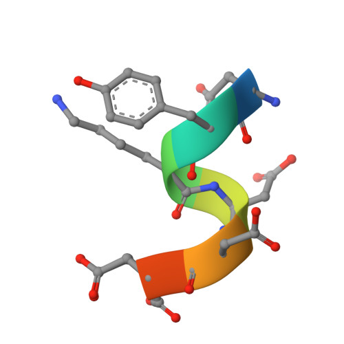
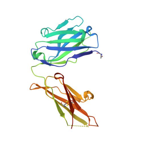
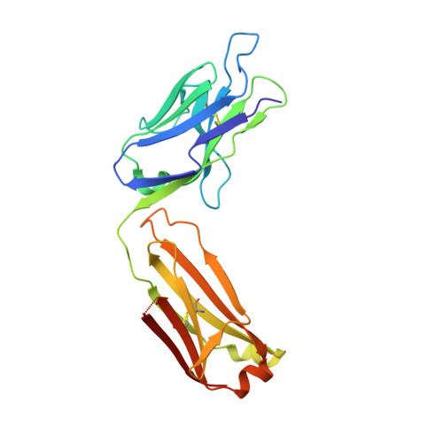
    The FLAG-tag/anti-FLAG system is a widely used biochemical tool for protein detection and purification. Anti-FLAG M2 is the most popular antibody against the FLAG-tag, due to its ease of use, versatility, and availability in pure form or as bead conjugate. M2 binds N-terminal, C-terminal and internal FLAG-tags and binding is calcium-independent, but the molecular basis for the FLAG-tag specificity and recognition remains unresolved. Here we present an atomic resolution (1.17 Å) structure of the FLAG peptide in complex with the Fab of anti-FLAG M2, revealing key binding determinants. Five of the eight FLAG peptide residues form direct interactions with paratope residues. The FLAG peptide adopts a 3 10 helix conformation in complex with the Fab. These structural insights allowed us to rationally introduce point mutations on both the peptide and antibody side. We tested these by surface plasmon resonance, leading us to propose a shorter yet equally binding version of the FLAG-tag for the M2 antibody.


  • Organizational Affiliation

    Structural Biochemistry, Bijvoet Centre for Biomolecular Research, Department of Chemistry, Faculty of Science, Utrecht University, Universiteitsweg 99, 3584 CG Utrecht, The Netherlands.


Macromolecules

Find similar proteins by:  Sequence   |   3D Structure  

Entity ID: 1
MoleculeChains Sequence LengthOrganismDetailsImage
FLAG-tagA [auth F]8synthetic constructMutation(s): 0 
Sequence Annotations
Expand
  • Reference Sequence
Find similar proteins by:  (by identity cutoff)  |  3D Structure
Entity ID: 2
MoleculeChains Sequence LengthOrganismDetailsImage
anti-FLAG M2 heavy chainB [auth H]228Mus musculusMutation(s): 0 
Entity Groups  
Sequence Clusters30% Identity50% Identity70% Identity90% Identity95% Identity100% Identity
Sequence Annotations
Expand
  • Reference Sequence
Find similar proteins by:  (by identity cutoff)  |  3D Structure
Entity ID: 3
MoleculeChains Sequence LengthOrganismDetailsImage
anti-FLAG M2 light chainC [auth L]219Mus musculusMutation(s): 0 
Entity Groups  
Sequence Clusters30% Identity50% Identity70% Identity90% Identity95% Identity100% Identity
Sequence Annotations
Expand
  • Reference Sequence
Small Molecules
Modified Residues  1 Unique
IDChains TypeFormula2D DiagramParent
PCA
Query on PCA
B [auth H]L-PEPTIDE LINKINGC5 H7 N O3GLN
Experimental Data & Validation

Experimental Data

  • Method: X-RAY DIFFRACTION
  • Resolution: 1.16 Å
  • R-Value Free: 0.185 
  • R-Value Work: 0.158 
  • Space Group: P 21 21 2
Unit Cell:
Length ( Å )Angle ( ˚ )
a = 87.432α = 90
b = 134.621β = 90
c = 41.798γ = 90
Software Package:
Software NamePurpose
REFMACrefinement
autoPROCdata reduction
STARANISOdata scaling
PHASERphasing

Structure Validation

View Full Validation Report


Entry History & Funding Information

Deposition Data


Funding OrganizationLocationGrant Number
Netherlands Organisation for Scientific Research (NWO)Netherlands--

Revision History  (Full details and data files)

  • Version 1.0: 2024-04-03
    Type: Initial release
  • Version 1.1: 2024-06-19
    Changes: Database references