A Multidisciplinary Structural Approach to the Identification of the Haemophilus influenzae Type b Capsular Polysaccharide Protective Epitope.
Nonne, F., Iacono, L.D., Bertuzzi, S., Unione, L., Proietti, D., Norais, N., Margarit, I., Adamo, R., Jimenez-Barbero, J., Carboni, F., Romano, M.R.(2024) ACS Cent Sci 10: 978-987
- PubMed: 38799664
- DOI: https://doi.org/10.1021/acscentsci.3c01515
- Primary Citation of Related Structures:
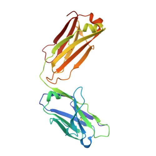
8RDA, 8RDF - PubMed Abstract:
Glycoconjugate vaccines so far licensed are generally composed of a native or size-reduced capsular polysaccharide conjugated to carrier proteins. Detailed information on the structural requirements necessary for CPS recognition is becoming the key to accelerating the development of next-generation improved glycoconjugate vaccines. Structural glycobiology studies using oligosaccharides (OS) complexed with functional monoclonal antibodies represent a powerful tool for gaining information on CPS immunological determinants at the atomic level. Herein, the minimal structural epitope of Haemophilus influenzae type b (Hib) CPS recognized by a functional human monoclonal antibody (hmAb) is reported. Short and well-defined Hib oligosaccharides originating from the depolymerization of the native CPS have been used to elucidate saccharide-mAb interactions by using a multidisciplinary approach combining surface plasmon resonance (SPR), saturation transfer difference-nanomagnetic resonance (STD-NMR), and X-ray crystallography. Our study demonstrates that the minimal structural epitope of Hib is comprised within two repeating units (RUs) where ribose and ribitol are directly engaged in the hmAb interaction, and the binding pocket fully accommodates two RUs without any additional involvement of a third one. Understanding saccharide antigen structural characteristics can provide the basis for the design of innovative glycoconjugate vaccines based on alternative technologies, such as synthetic or enzymatic approaches.
Organizational Affiliation:
GSK Vaccines Institute for Global Health, 53100 Siena, Italy.