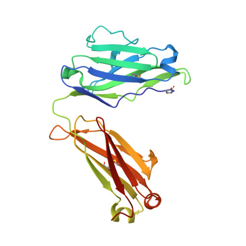
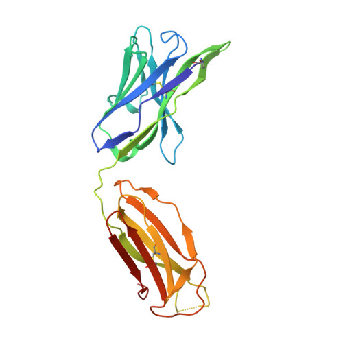
8OJS

Crystal structure of the human IgD Fab - structure Fab1


Experimental Data Snapshot

  • Method: X-RAY DIFFRACTION
  • Resolution: 2.75 Å
  • R-Value Free: 0.256 
  • R-Value Work: 0.216 
  • R-Value Observed: 0.218 

Starting Model: experimental
View more details

wwPDB Validation   3D Report Full Report


This is version 1.1 of the entry. See complete history


Literature

Crystal structures of the human IgD Fab reveal insights into C H 1 domain diversity.

Davies, A.M.Beavil, R.L.Barbolov, M.Sandhar, B.S.Gould, H.J.Beavil, A.J.Sutton, B.J.McDonnell, J.M.

(2023) Mol Immunol 159: 28-37

  • DOI: https://doi.org/10.1016/j.molimm.2023.05.006
  • Primary Citation of Related Structures:  
    8OJS, 8OJT, 8OJU, 8OJV

  • PubMed Abstract: 

    Antibodies of the IgD isotype remain the least well characterized of the mammalian immunoglobulin isotypes. Here we report three-dimensional structures for the Fab region of IgD, based on four different crystal structures, at resolutions of 1.45-2.75 Å. These IgD Fab crystals provide the first high-resolution views of the unique Cδ1 domain. Structural comparisons identify regions of conformational diversity within the Cδ1 domain, as well as among the homologous domains of Cα1, Cγ1 and Cμ1. The IgD Fab structure also possesses a unique conformation of the upper hinge region, which may contribute to the overall disposition of the very long linker sequence between the Fab and Fc regions found in human IgD. Structural similarities observed between IgD and IgG, and differences with IgA and IgM, are consistent with predicted evolutionary relationships for the mammalian antibody isotypes.


  • Organizational Affiliation

    King's College London, Randall Centre for Cell and Molecular Biophysics, New Hunt's House, London SE1 1UL, United Kingdom.


Macromolecules
Find similar proteins by:  (by identity cutoff)  |  3D Structure
Entity ID: 1
MoleculeChains Sequence LengthOrganismDetailsImage
Human IgD Fab light chainA,
C [auth L]
217Homo sapiensMutation(s): 0 
Entity Groups  
Sequence Clusters30% Identity50% Identity70% Identity90% Identity95% Identity100% Identity
Sequence Annotations
Expand
  • Reference Sequence
Find similar proteins by:  (by identity cutoff)  |  3D Structure
Entity ID: 2
MoleculeChains Sequence LengthOrganismDetailsImage
Human IgD Fab heavy chainB,
D [auth H]
227Homo sapiensMutation(s): 0 
Entity Groups  
Sequence Clusters30% Identity50% Identity70% Identity90% Identity95% Identity100% Identity
Sequence Annotations
Expand
  • Reference Sequence
Small Molecules
Modified Residues  1 Unique
IDChains TypeFormula2D DiagramParent
PCA
Query on PCA
A,
C [auth L]
L-PEPTIDE LINKINGC5 H7 N O3GLN
Experimental Data & Validation

Experimental Data

  • Method: X-RAY DIFFRACTION
  • Resolution: 2.75 Å
  • R-Value Free: 0.256 
  • R-Value Work: 0.216 
  • R-Value Observed: 0.218 
  • Space Group: P 1
Unit Cell:
Length ( Å )Angle ( ˚ )
a = 43.534α = 92.52
b = 71.789β = 91.16
c = 90.861γ = 105.33
Software Package:
Software NamePurpose
PHENIXrefinement
Aimlessdata scaling
DIALSdata reduction
PHASERphasing

Structure Validation

View Full Validation Report


Entry History & Funding Information

Deposition Data


Funding OrganizationLocationGrant Number
Medical Research Council (MRC, United Kingdom)United KingdomMR/V010557/1

Revision History  (Full details and data files)

  • Version 1.0: 2023-06-14
    Type: Initial release
  • Version 1.1: 2024-10-16
    Changes: Data collection, Structure summary