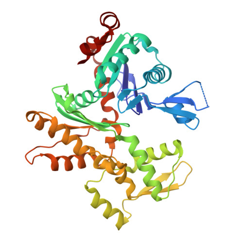
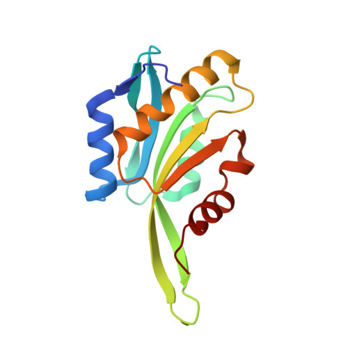
8OH4

Subtomogram averaging structure of cofilactin filament inside microtubule lumen of Drosophila S2 cell protrusion.


Experimental Data Snapshot

  • Method: ELECTRON MICROSCOPY
  • Resolution: 16.5 Å
  • Aggregation State: FILAMENT 
  • Reconstruction Method: SUBTOMOGRAM AVERAGING 

wwPDB Validation   3D Report Full Report


This is version 1.1 of the entry. See complete history


Literature

CryoET shows cofilactin filaments inside the microtubule lumen.

Santos, C.V.Rogers, S.L.Carter, A.P.

(2023) bioRxiv 

  • DOI: https://doi.org/10.1101/2023.03.31.535077
  • Primary Citation of Related Structures:  
    8OH4

  • PubMed Abstract: 

    Cytoplasmic microtubules are tubular polymers that can harbor small proteins or filaments inside their lumen. The identity of these objects and what causes their accumulation has not been conclusively established. Here, we used cryogenic electron tomography (cryoET) of Drosophila S2 cell protrusions and found filaments inside the microtubule lumen, which resemble those reported recently in human HAP1 cells. The frequency of these filaments increased upon inhibition of the sarco/endoplasmic reticulum Ca 2+ ATPase (SERCA) with the small-molecule drug thapsigargin. Subtomogram averaging showed that the luminal filaments adopt a helical structure reminiscent of cofilin-bound actin (cofilactin). Consistent with this, cofilin was activated in cells under the same conditions that increased luminal filament occurrence. Furthermore, RNAi knock-down of cofilin reduced the frequency of luminal filaments with cofilactin morphology. These results suggest that cofilin activation stimulates its accumulation on actin filaments inside the microtubule lumen.


  • Organizational Affiliation

    MRC Laboratory of Molecular Biology, Francis Crick Ave, Cambridge, CB2 0QH, UK.


Macromolecules
Find similar proteins by:  (by identity cutoff)  |  3D Structure
Entity ID: 1
MoleculeChains Sequence LengthOrganismDetailsImage
Actin-5C370DrosophilaMutation(s): 0 
EC: 3.6.4
UniProt
Find proteins for P10987 (Drosophila melanogaster)
Explore P10987 
Go to UniProtKB:  P10987
Entity Groups  
Sequence Clusters30% Identity50% Identity70% Identity90% Identity95% Identity100% Identity
UniProt GroupP10987
Sequence Annotations
Expand
  • Reference Sequence
Find similar proteins by:  (by identity cutoff)  |  3D Structure
Entity ID: 2
MoleculeChains Sequence LengthOrganismDetailsImage
Cofilin/actin-depolymerizing factor homolog148DrosophilaMutation(s): 0 
UniProt
Find proteins for P45594 (Drosophila melanogaster)
Explore P45594 
Go to UniProtKB:  P45594
Entity Groups  
Sequence Clusters30% Identity50% Identity70% Identity90% Identity95% Identity100% Identity
UniProt GroupP45594
Sequence Annotations
Expand
  • Reference Sequence
Experimental Data & Validation

Experimental Data

  • Method: ELECTRON MICROSCOPY
  • Resolution: 16.5 Å
  • Aggregation State: FILAMENT 
  • Reconstruction Method: SUBTOMOGRAM AVERAGING 
EM Software:
TaskSoftware PackageVersion
RECONSTRUCTIONRELION3.1

Structure Validation

View Full Validation Report


Entry History & Funding Information

Deposition Data


Funding OrganizationLocationGrant Number
Medical Research Council (MRC, United Kingdom)United KingdomMC_UP_A025_1011
Wellcome TrustUnited Kingdom210711/Z/18/Z

Revision History  (Full details and data files)

  • Version 1.0: 2023-05-10
    Type: Initial release
  • Version 1.1: 2024-07-24
    Changes: Data collection