8GUE

Crystal Structure of narbomycin-bound cytochrome P450 PikC with the unnatural amino acid p-Acetyl-L-Phenylalanine incorporated at position 238


Experimental Data Snapshot

  • Method: X-RAY DIFFRACTION
  • Resolution: 1.90 Å
  • R-Value Free: 0.250 
  • R-Value Work: 0.201 
  • R-Value Observed: 0.203 

Starting Model: experimental
View more details

wwPDB Validation   3D Report Full Report


Ligand Structure Quality Assessment 


This is version 2.1 of the entry. See complete history


Literature

Unnatural activities and mechanistic insights of cytochrome P450 PikC gained from site-specific mutagenesis by non-canonical amino acids.

Pan, Y.Li, G.Liu, R.Guo, J.Liu, Y.Liu, M.Zhang, X.Chi, L.Xu, K.Wu, R.Zhang, Y.Li, Y.Gao, X.Li, S.

(2023) Nat Commun 14: 1669-1669

  • DOI: https://doi.org/10.1038/s41467-023-37288-0
  • Primary Citation of Related Structures:  
    8GUE

  • PubMed Abstract: 

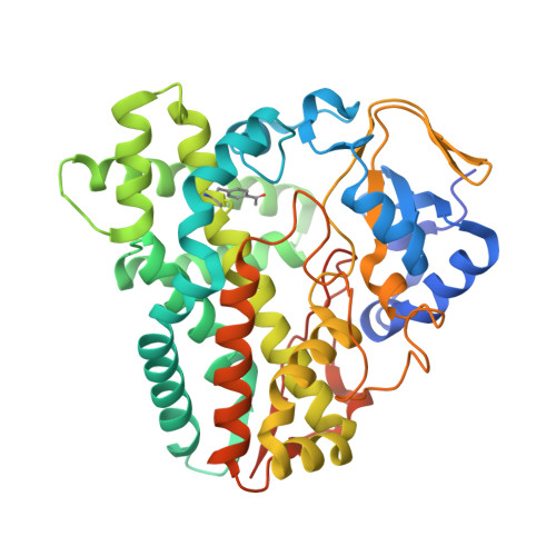
    Cytochrome P450 enzymes play important roles in the biosynthesis of macrolide antibiotics by mediating a vast variety of regio- and stereoselective oxidative modifications, thus improving their chemical diversity, biological activities, and pharmaceutical properties. Tremendous efforts have been made on engineering the reactivity and selectivity of these useful biocatalysts. However, the 20 proteinogenic amino acids cannot always satisfy the requirement of site-directed/random mutagenesis and rational protein design of P450 enzymes. To address this issue, herein, we practice the semi-rational non-canonical amino acid mutagenesis for the pikromycin biosynthetic P450 enzyme PikC, which recognizes its native macrolide substrates with a 12- or 14-membered ring macrolactone linked to a deoxyamino sugar through a unique sugar-anchoring mechanism. Based on a semi-rationally designed substrate binding strategy, non-canonical amino acid mutagenesis at the His238 position enables the unnatural activities of several PikC mutants towards the macrolactone precursors without any sugar appendix. With the aglycone hydroxylating activities, the pikromycin biosynthetic pathway is rewired by the representative mutant PikC H238pAcF carrying a p-acetylphenylalanine residue at the His238 position and a promiscuous glycosyltransferase. Moreover, structural analysis of substrate-free and three different enzyme-substrate complexes of PikC H238pAcF provides significant mechanistic insights into the substrate binding and catalytic selectivity of this paradigm biosynthetic P450 enzyme.


  • Organizational Affiliation

    State Key Laboratory of Microbial Technology, Shandong University, Qingdao, Shandong, 266237, China.


Macromolecules
Find similar proteins by:  (by identity cutoff)  |  3D Structure
Entity ID: 1
MoleculeChains Sequence LengthOrganismDetailsImage
Cytochrome P450 monooxygenase PikC
A, B
424Streptomyces venezuelaeMutation(s): 0 
Gene Names: pikCpicK
EC: 1.14.15.33
UniProt
Find proteins for O87605 (Streptomyces venezuelae)
Explore O87605 
Go to UniProtKB:  O87605
Entity Groups  
Sequence Clusters30% Identity50% Identity70% Identity90% Identity95% Identity100% Identity
UniProt GroupO87605
Sequence Annotations
Expand
  • Reference Sequence
Small Molecules
Ligands 6 Unique
IDChains Name / Formula / InChI Key2D Diagram3D Interactions
HEM (Subject of Investigation/LOI)
Query on HEM

Download Ideal Coordinates CCD File 
C [auth A],
N [auth B]
PROTOPORPHYRIN IX CONTAINING FE
C34 H32 Fe N4 O4
KABFMIBPWCXCRK-RGGAHWMASA-L
NRB (Subject of Investigation/LOI)
Query on NRB

Download Ideal Coordinates CCD File 
D [auth A],
O [auth B]
NARBOMYCIN
C28 H47 N O7
OXFYAOOMMKGGAI-ZMIJTAGNSA-N
TRS
Query on TRS

Download Ideal Coordinates CCD File 
L [auth A]2-AMINO-2-HYDROXYMETHYL-PROPANE-1,3-DIOL
C4 H12 N O3
LENZDBCJOHFCAS-UHFFFAOYSA-O
PEG
Query on PEG

Download Ideal Coordinates CCD File 
M [auth A],
V [auth B]
DI(HYDROXYETHYL)ETHER
C4 H10 O3
MTHSVFCYNBDYFN-UHFFFAOYSA-N
SO4
Query on SO4

Download Ideal Coordinates CCD File 
E [auth A]
F [auth A]
G [auth A]
H [auth A]
I [auth A]
E [auth A],
F [auth A],
G [auth A],
H [auth A],
I [auth A],
J [auth A],
K [auth A],
P [auth B],
Q [auth B],
R [auth B],
T [auth B]
SULFATE ION
O4 S
QAOWNCQODCNURD-UHFFFAOYSA-L
EDO
Query on EDO

Download Ideal Coordinates CCD File 
S [auth B],
U [auth B]
1,2-ETHANEDIOL
C2 H6 O2
LYCAIKOWRPUZTN-UHFFFAOYSA-N
Modified Residues  1 Unique
IDChains TypeFormula2D DiagramParent
4AF
Query on 4AF
A, B
L-PEPTIDE LINKINGC11 H13 N O3PHE
Experimental Data & Validation

Experimental Data

  • Method: X-RAY DIFFRACTION
  • Resolution: 1.90 Å
  • R-Value Free: 0.250 
  • R-Value Work: 0.201 
  • R-Value Observed: 0.203 
  • Space Group: P 21 21 21
Unit Cell:
Length ( Å )Angle ( ˚ )
a = 59.465α = 90
b = 97.818β = 90
c = 141.075γ = 90
Software Package:
Software NamePurpose
PHENIXrefinement
HKL-3000data reduction
HKL-3000data scaling
PHENIXphasing

Structure Validation

View Full Validation Report


Ligand Structure Quality Assessment 


Entry History & Funding Information

Deposition Data


Funding OrganizationLocationGrant Number
National Natural Science Foundation of China (NSFC)China32000039
National Natural Science Foundation of China (NSFC)China32025001
National Natural Science Foundation of China (NSFC)China31872729

Revision History  (Full details and data files)

  • Version 1.0: 2023-02-15
    Type: Initial release
  • Version 1.1: 2023-09-20
    Changes: Data collection, Database references
  • Version 2.0: 2023-11-15
    Changes: Atomic model, Data collection, Derived calculations
  • Version 2.1: 2023-11-29
    Changes: Refinement description