8G2O

Fab structure - Anti-ApoE-7C11 antibody


Experimental Data Snapshot

  • Method: X-RAY DIFFRACTION
  • Resolution: 1.70 Å
  • R-Value Free: 0.204 
  • R-Value Work: 0.186 
  • R-Value Observed: 0.187 

Starting Model: experimental
View more details

wwPDB Validation   3D Report Full Report


Ligand Structure Quality Assessment 


This is version 1.1 of the entry. See complete history


Literature

APOE Christchurch-mimetic therapeutic antibody reduces APOE-mediated toxicity and tau phosphorylation.

Marino, C.Perez-Corredor, P.O'Hare, M.Heuer, A.Chmielewska, N.Gordon, H.Chandrahas, A.S.Gonzalez-Buendia, L.Delgado-Tirado, S.Doan, T.H.Vanderleest, T.E.Arevalo-Alquichire, S.Obar, R.A.Ortiz-Cordero, C.Villegas, A.Sepulveda-Falla, D.Kim, L.A.Lopera, F.Mahley, R.Huang, Y.Quiroz, Y.T.Arboleda-Velasquez, J.F.

(2024) Alzheimers Dement 20: 819-836

  • DOI: https://doi.org/10.1002/alz.13436
  • Primary Citation of Related Structures:  
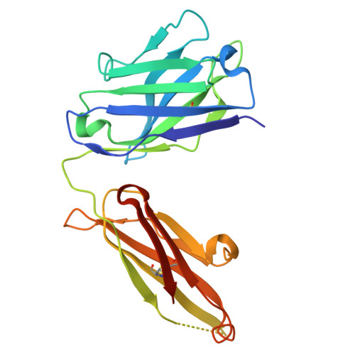
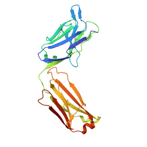
    8G2O

  • PubMed Abstract: 

    We discovered that the APOE3 Christchurch (APOE3Ch) variant may provide resistance to Alzheimer's disease (AD). This resistance may be due to reduced pathological interactions between ApoE3Ch and heparan sulfate proteoglycans (HSPGs). We developed and characterized the binding, structure, and preclinical efficacy of novel antibodies targeting human ApoE-HSPG interactions. We found that one of these antibodies, called 7C11, preferentially bound ApoE4, a major risk factor for sporadic AD, and disrupts heparin-ApoE4 interactions. We also determined the crystal structure of a Fab fragment of 7C11 and used computer modeling to predict how it would bind to ApoE. When we tested 7C11 in mouse models, we found that it reduced recombinant ApoE-induced tau pathology in the retina of MAPT*P301S mice and curbed pTau S396 phosphorylation in brains of systemically treated APOE4 knock-in mice. Targeting ApoE-HSPG interactions using 7C11 antibody may be a promising approach to developing new therapies for AD.


  • Organizational Affiliation

    Schepens Eye Research Institute of Mass Eye and Ear and Department of Ophthalmology at Harvard Medical School, Boston, Massachusetts, USA.


Macromolecules
Find similar proteins by:  (by identity cutoff)  |  3D Structure
Entity ID: 1
MoleculeChains Sequence LengthOrganismDetailsImage
anti-ApoE-7C11 heavy chainA [auth H]225Homo sapiensMutation(s): 0 
Entity Groups  
Sequence Clusters30% Identity50% Identity70% Identity90% Identity95% Identity100% Identity
Sequence Annotations
Expand
  • Reference Sequence
Find similar proteins by:  (by identity cutoff)  |  3D Structure
Entity ID: 2
MoleculeChains Sequence LengthOrganismDetailsImage
anti-ApoE-7C11 light chainB [auth L]218Homo sapiensMutation(s): 0 
Entity Groups  
Sequence Clusters30% Identity50% Identity70% Identity90% Identity95% Identity100% Identity
Glycosylation
Glycosylation Sites: 1
Sequence Annotations
Expand
  • Reference Sequence
Small Molecules
Ligands 1 Unique
IDChains Name / Formula / InChI Key2D Diagram3D Interactions
NAG (Subject of Investigation/LOI)
Query on NAG

Download Ideal Coordinates CCD File 
C [auth L]2-acetamido-2-deoxy-beta-D-glucopyranose
C8 H15 N O6
OVRNDRQMDRJTHS-FMDGEEDCSA-N
Experimental Data & Validation

Experimental Data

  • Method: X-RAY DIFFRACTION
  • Resolution: 1.70 Å
  • R-Value Free: 0.204 
  • R-Value Work: 0.186 
  • R-Value Observed: 0.187 
  • Space Group: P 21 21 2
Unit Cell:
Length ( Å )Angle ( ˚ )
a = 92.7α = 90
b = 62.27β = 90
c = 82.14γ = 90
Software Package:
Software NamePurpose
PHENIXrefinement
XDSdata reduction
XDSdata scaling
MOLREPphasing

Structure Validation

View Full Validation Report


Ligand Structure Quality Assessment 


Entry History & Funding Information

Deposition Data


Funding OrganizationLocationGrant Number
National Institutes of Health/National Institute of Neurological Disorders and Stroke (NIH/NINDS)United StatesNS100121
National Institutes of Health/National Institute of Neurological Disorders and Stroke (NIH/NINDS)United StatesNS110048
Other privateUnited States2020A011571

Revision History  (Full details and data files)

  • Version 1.0: 2023-10-18
    Type: Initial release
  • Version 1.1: 2024-02-28
    Changes: Database references