8FSL

Human Mesothelin bound to a neutralizing VH domain antibody


Experimental Data Snapshot

  • Method: X-RAY DIFFRACTION
  • Resolution: 2.90 Å
  • R-Value Free: 0.254 
  • R-Value Work: 0.238 
  • R-Value Observed: 0.239 

Starting Model: experimental
View more details

wwPDB Validation   3D Report Full Report


This is version 1.0 of the entry. See complete history


Literature

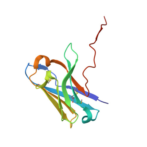
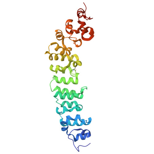
Preclinical assessment of a novel human antibody VH domain targeting mesothelin as an antibody-drug conjugate.

Sun, Z.Chu, X.Adams, C.Ilina, T.V.Guerrero, M.Lin, G.Chen, C.Jelev, D.Ishima, R.Li, W.Mellors, J.W.Calero, G.Dimitrov, D.S.

(2023) Mol Ther Oncolytics 31: 100726-100726

  • DOI: https://doi.org/10.1016/j.omto.2023.09.002
  • Primary Citation of Related Structures:  
    8FSL

  • PubMed Abstract: 

    Mesothelin (MSLN) has been a validated tumor-associated antigen target for several solid tumors for over a decade, making it an attractive option for therapeutic interventions. Novel antibodies with high affinity and better therapeutic properties are needed. In the current study, we have isolated and characterized a novel heavy chain variable (VH) domain 3C9 from a large-size human immunoglobulin VH domain library. 3C9 exhibited high affinity (KD [dissociation constant] <3 nM) and binding specificity in a membrane proteome array (MPA). In a mouse xenograft model, 3C9 fused to human IgG1 Fc was detected at tumor sites as early as 8 h post-infusion and remained at the site for over 10 days. Furthermore, 3C9 fused to a human Fc domain drug conjugate effectively inhibited MSLN-positive tumor growth in a mouse xenograft model. The X-ray crystal structure of full-length MSLN in complex with 3C9 reveals interaction of the 3C9 domains with two distinctive residue patches on the MSLN surface. This newly discovered VH antibody domain has a high potential as a therapeutic candidate for MSLN-expressing cancers.


  • Organizational Affiliation

    Center for Antibody Therapeutics, Division of Infectious Diseases, Department of Medicine, University of Pittsburgh School of Medicine, 3550 Terrace Street, Pittsburgh, PA 15261, USA.


Macromolecules
Find similar proteins by:  (by identity cutoff)  |  3D Structure
Entity ID: 1
MoleculeChains Sequence LengthOrganismDetailsImage
VH domainA,
B [auth C],
C [auth D],
F [auth B]
116Escherichia coliMutation(s): 0 
Entity Groups  
Sequence Clusters30% Identity50% Identity70% Identity90% Identity95% Identity100% Identity
Sequence Annotations
Expand
  • Reference Sequence
Find similar proteins by:  (by identity cutoff)  |  3D Structure
Entity ID: 2
MoleculeChains Sequence LengthOrganismDetailsImage
MesothelinD [auth E],
E [auth F]
285Insect expression vector pBlueBacmsGCA1HisMutation(s): 0 
Gene Names: MSLNMPF
UniProt & NIH Common Fund Data Resources
Find proteins for Q13421 (Homo sapiens)
Explore Q13421 
Go to UniProtKB:  Q13421
PHAROS:  Q13421
GTEx:  ENSG00000102854 
Entity Groups  
Sequence Clusters30% Identity50% Identity70% Identity90% Identity95% Identity100% Identity
UniProt GroupQ13421
Sequence Annotations
Expand
  • Reference Sequence
Small Molecules
Modified Residues  1 Unique
IDChains TypeFormula2D DiagramParent
MSE
Query on MSE
A,
B [auth C],
C [auth D],
F [auth B]
L-PEPTIDE LINKINGC5 H11 N O2 SeMET
Experimental Data & Validation

Experimental Data

  • Method: X-RAY DIFFRACTION
  • Resolution: 2.90 Å
  • R-Value Free: 0.254 
  • R-Value Work: 0.238 
  • R-Value Observed: 0.239 
  • Space Group: P 43 2 2
Unit Cell:
Length ( Å )Angle ( ˚ )
a = 114.116α = 90
b = 114.116β = 90
c = 340.74γ = 90
Software Package:
Software NamePurpose
PHENIXrefinement
PHENIXrefinement
XDSdata reduction
XDSdata scaling
CRANK2phasing

Structure Validation

View Full Validation Report


Entry History & Funding Information

Deposition Data

  • Released Date: 2024-05-29 
  • Deposition Author(s): Calero, G.

Funding OrganizationLocationGrant Number
Other privateNA

Revision History  (Full details and data files)

  • Version 1.0: 2024-05-29
    Type: Initial release