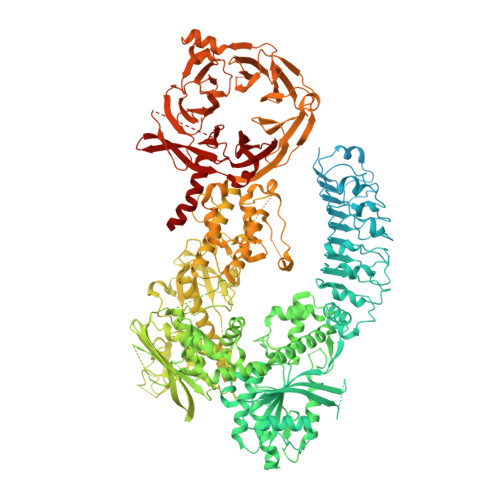
8E04

Structure of monomeric LRRK1


Experimental Data Snapshot

  • Method: ELECTRON MICROSCOPY
  • Resolution: 3.80 Å
  • Aggregation State: PARTICLE 
  • Reconstruction Method: SINGLE PARTICLE 

wwPDB Validation   3D Report Full Report


This is version 1.2 of the entry. See complete history


Literature

Structure of LRRK1 and mechanisms of autoinhibition and activation.

Reimer, J.M.Dickey, A.M.Lin, Y.X.Abrisch, R.G.Mathea, S.Chatterjee, D.Fay, E.J.Knapp, S.Daugherty, M.D.Reck-Peterson, S.L.Leschziner, A.E.

(2023) Nat Struct Mol Biol 30: 1735-1745

  • DOI: https://doi.org/10.1038/s41594-023-01109-1
  • Primary Citation of Related Structures:  
    8E04, 8E05, 8E06

  • PubMed Abstract: 

    Leucine Rich Repeat Kinase 1 and 2 (LRRK1 and LRRK2) are homologs in the ROCO family of proteins in humans. Despite their shared domain architecture and involvement in intracellular trafficking, their disease associations are strikingly different: LRRK2 is involved in familial Parkinson's disease while LRRK1 is linked to bone diseases. Furthermore, Parkinson's disease-linked mutations in LRRK2 are typically autosomal dominant gain-of-function while those in LRRK1 are autosomal recessive loss-of-function. Here, to understand these differences, we solved cryo-EM structures of LRRK1 in its monomeric and dimeric forms. Both differ from the corresponding LRRK2 structures. Unlike LRRK2, which is sterically autoinhibited as a monomer, LRRK1 is sterically autoinhibited in a dimer-dependent manner. LRRK1 has an additional level of autoinhibition that prevents activation of the kinase and is absent in LRRK2. Finally, we place the structural signatures of LRRK1 and LRRK2 in the context of the evolution of the LRRK family of proteins.


  • Organizational Affiliation

    Department of Cellular and Molecular Medicine, University of California San Diego, La Jolla, CA, USA.


Macromolecules
Find similar proteins by:  (by identity cutoff)  |  3D Structure
Entity ID: 1
MoleculeChains Sequence LengthOrganismDetailsImage
Leucine-rich repeat serine/threonine-protein kinase 11,996Homo sapiensMutation(s): 0 
Gene Names: LRRK1KIAA1790
EC: 2.7.11.1
UniProt & NIH Common Fund Data Resources
Find proteins for Q38SD2 (Homo sapiens)
Explore Q38SD2 
Go to UniProtKB:  Q38SD2
PHAROS:  Q38SD2
GTEx:  ENSG00000154237 
Entity Groups  
Sequence Clusters30% Identity50% Identity70% Identity90% Identity95% Identity100% Identity
UniProt GroupQ38SD2
Sequence Annotations
Expand
  • Reference Sequence
Small Molecules
Ligands 1 Unique
IDChains Name / Formula / InChI Key2D Diagram3D Interactions
GDP
Query on GDP

Download Ideal Coordinates CCD File 
B [auth A]GUANOSINE-5'-DIPHOSPHATE
C10 H15 N5 O11 P2
QGWNDRXFNXRZMB-UUOKFMHZSA-N
Experimental Data & Validation

Experimental Data

  • Method: ELECTRON MICROSCOPY
  • Resolution: 3.80 Å
  • Aggregation State: PARTICLE 
  • Reconstruction Method: SINGLE PARTICLE 

Structure Validation

View Full Validation Report


Entry History & Funding Information

Deposition Data


Funding OrganizationLocationGrant Number
Damon Runyon Cancer Research FoundationUnited StatesDRG-2370-19
Michael J. Fox FoundationUnited States18321
Other privateASAP-000519
National Institutes of Health/National Institute of General Medical Sciences (NIH/NIGMS)United StatesR01GM107214

Revision History  (Full details and data files)

  • Version 1.0: 2023-08-30
    Type: Initial release
  • Version 1.1: 2023-11-01
    Changes: Database references
  • Version 1.2: 2023-11-29
    Changes: Database references