8DHD

Neutron crystal structure of maltotetraose bound tmMBP


Experimental Data Snapshot

  • Method: X-RAY DIFFRACTION
  • Resolution: 1.70 Å
  • R-Value Free: 0.175 
  • R-Value Work: 0.141 
  • R-Value Observed: 0.143 

  • Method: NEUTRON DIFFRACTION
  • Resolution: 2.11 Å
  • R-Value Free: 0.272 
  • R-Value Work: 0.245 
  • R-Value Observed: 0.246 

wwPDB Validation   3D Report Full Report


This is version 1.2 of the entry. See complete history


Literature

Mapping periplasmic binding protein oligosaccharide recognition with neutron crystallography.

Shukla, S.Myles, D.A.Cuneo, M.J.

(2022) Sci Rep 12: 17647-17647

  • DOI: https://doi.org/10.1038/s41598-022-20542-8
  • Primary Citation of Related Structures:  
    8DHD

  • PubMed Abstract: 

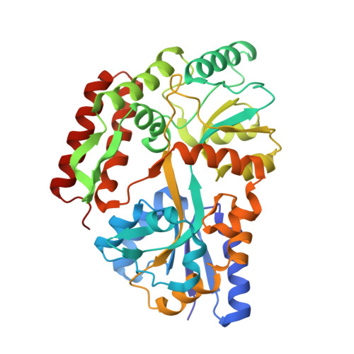
    Numerous studies have shown how periplasmic binding proteins (PBPs) bind substrates with exquisite specificity, even distinguishing between sugar epimers and anomers, or structurally similar ions. Yet, marked substrate promiscuity is also a feature encoded in some PBPs. Except for three sub-Ångström crystal structures, there are no reports of hydrogen atom positions in the remaining (> 1000) PBP structures. The previous X-ray crystal structure of the maltodextrin periplasmic-binding protein from Thermotoga maritima (tmMBP) complexed with oligosaccharide showed a large network of interconnected water molecules stretching from one end of the substrate binding pocket to the other. These water molecules are positioned to form multiple hydrogen bonds, as well as forming interactions between the protein and substrate. Here we present the neutron crystal structure of tmMBP to a resolution of 2.1 Å. This is the first neutron crystal structure from the PBP superfamily and here we unambiguously identify the nature and orientation of the hydrogen bonding and water-mediated interactions involved in stabilizing a tetrasaccharide in the binding site. More broadly, these results demonstrate the conserved intricate mechanisms that underlie substrate-specificity and affinity in PBPs.


  • Organizational Affiliation

    Neutron Scattering Division, Oak Ridge National Laboratory, Oak Ridge, TN, 37831, USA.


Macromolecules
Find similar proteins by:  (by identity cutoff)  |  3D Structure
Entity ID: 1
MoleculeChains Sequence LengthOrganismDetailsImage
maltose-binding protein MalE2383Thermotoga maritima MSB8Mutation(s): 0 
Gene Names: TM_1839
UniProt
Find proteins for Q9S5Y1 (Thermotoga maritima (strain ATCC 43589 / DSM 3109 / JCM 10099 / NBRC 100826 / MSB8))
Explore Q9S5Y1 
Go to UniProtKB:  Q9S5Y1
Entity Groups  
Sequence Clusters30% Identity50% Identity70% Identity90% Identity95% Identity100% Identity
UniProt GroupQ9S5Y1
Sequence Annotations
Expand
  • Reference Sequence
Oligosaccharides

Help

Entity ID: 2
MoleculeChains Length2D Diagram Glycosylation3D Interactions
alpha-D-glucopyranose-(1-4)-alpha-D-glucopyranose-(1-4)-alpha-D-glucopyranose-(1-4)-alpha-D-glucopyranose
B
4N/A
Glycosylation Resources
GlyTouCan:  G87171PZ
GlyCosmos:  G87171PZ
GlyGen:  G87171PZ
Biologically Interesting Molecules (External Reference) 1 Unique
Experimental Data & Validation

Experimental Data

  • Method: X-RAY DIFFRACTION
  • Resolution: 1.70 Å
  • R-Value Free: 0.175 
  • R-Value Work: 0.141 
  • R-Value Observed: 0.143 
  • Space Group: P 1 21 1
  • Method: NEUTRON DIFFRACTION
  • Resolution: 2.11 Å
  • R-Value Free: 0.272 
  • R-Value Work: 0.245 
  • R-Value Observed: 0.246 
  • Space Group: P 1 21 1
Unit Cell:
Length ( Å )Angle ( ˚ )
a = 35.915α = 90
b = 56.34β = 94.25
c = 90.001γ = 90
Software Package:
Software NamePurpose
PHENIXrefinement
PDB_EXTRACTdata extraction
CrysalisProdata reduction
LAUENORMdata scaling
PHASERphasing

Structure Validation

View Full Validation Report


Entry History & Funding Information

Deposition Data


Funding OrganizationLocationGrant Number
Not funded--

Revision History  (Full details and data files)

  • Version 1.0: 2022-10-12
    Type: Initial release
  • Version 1.1: 2022-11-02
    Changes: Database references
  • Version 1.2: 2023-10-25
    Changes: Data collection, Refinement description