8D5B

Crystal structure of human METTL1 in complex with SAH

  • Classification: TRANSFERASE
  • Organism(s): Homo sapiens
  • Expression System: Escherichia coli BL21(DE3)
  • Mutation(s): No 

  • Deposited: 2022-06-04 Released: 2023-01-11 
  • Deposition Author(s): Raj, R., Babu, K., Nam, Y.
  • Funding Organization(s): National Institutes of Health/National Institute of General Medical Sciences (NIH/NIGMS), Cancer Prevention and Research Institute of Texas (CPRIT), Welch Foundation

Experimental Data Snapshot

  • Method: X-RAY DIFFRACTION
  • Resolution: 1.93 Å
  • R-Value Free: 0.212 
  • R-Value Work: 0.181 
  • R-Value Observed: 0.182 

Starting Model: experimental
View more details

wwPDB Validation   3D Report Full Report


Ligand Structure Quality Assessment 


This is version 1.2 of the entry. See complete history


Literature

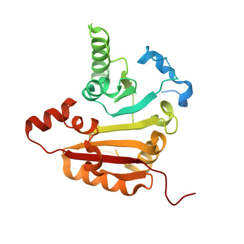
Structures and mechanisms of tRNA methylation by METTL1-WDR4.

Ruiz-Arroyo, V.M.Raj, R.Babu, K.Onolbaatar, O.Roberts, P.H.Nam, Y.

(2023) Nature 613: 383-390

  • DOI: https://doi.org/10.1038/s41586-022-05565-5
  • Primary Citation of Related Structures:  
    8D58, 8D59, 8D5B, 8D9K, 8D9L, 8EG0

  • PubMed Abstract: 

    Specific, regulated modification of RNAs is important for proper gene expression 1,2 . tRNAs are rich with various chemical modifications that affect their stability and function 3,4 . 7-Methylguanosine (m 7 G) at tRNA position 46 is a conserved modification that modulates steady-state tRNA levels to affect cell growth 5,6 . The METTL1-WDR4 complex generates m 7 G46 in humans, and dysregulation of METTL1-WDR4 has been linked to brain malformation and multiple cancers 7-22 . Here we show how METTL1 and WDR4 cooperate to recognize RNA substrates and catalyse methylation. A crystal structure of METTL1-WDR4 and cryo-electron microscopy structures of METTL1-WDR4-tRNA show that the composite protein surface recognizes the tRNA elbow through shape complementarity. The cryo-electron microscopy structures of METTL1-WDR4-tRNA with S-adenosylmethionine or S-adenosylhomocysteine along with METTL1 crystal structures provide additional insights into the catalytic mechanism by revealing the active site in multiple states. The METTL1 N terminus couples cofactor binding with conformational changes in the tRNA, the catalytic loop and the WDR4 C terminus, acting as the switch to activate m 7 G methylation. Thus, our structural models explain how post-translational modifications of the METTL1 N terminus can regulate methylation. Together, our work elucidates the core and regulatory mechanisms underlying m 7 G modification by METTL1, providing the framework to understand its contribution to biology and disease.


  • Organizational Affiliation

    Department of Biochemistry, University of Texas Southwestern Medical Center, Dallas, TX, USA.


Macromolecules
Find similar proteins by:  (by identity cutoff)  |  3D Structure
Entity ID: 1
MoleculeChains Sequence LengthOrganismDetailsImage
tRNA (guanine-N(7)-)-methyltransferase262Homo sapiensMutation(s): 0 
Gene Names: METTL1C12orf1
EC: 2.1.1.33 (PDB Primary Data), 2.1.1 (PDB Primary Data)
UniProt & NIH Common Fund Data Resources
Find proteins for Q9UBP6 (Homo sapiens)
Explore Q9UBP6 
Go to UniProtKB:  Q9UBP6
PHAROS:  Q9UBP6
GTEx:  ENSG00000037897 
Entity Groups  
Sequence Clusters30% Identity50% Identity70% Identity90% Identity95% Identity100% Identity
UniProt GroupQ9UBP6
Sequence Annotations
Expand
  • Reference Sequence
Small Molecules
Ligands 5 Unique
IDChains Name / Formula / InChI Key2D Diagram3D Interactions
SAH (Subject of Investigation/LOI)
Query on SAH

Download Ideal Coordinates CCD File 
B [auth A]S-ADENOSYL-L-HOMOCYSTEINE
C14 H20 N6 O5 S
ZJUKTBDSGOFHSH-WFMPWKQPSA-N
CAC
Query on CAC

Download Ideal Coordinates CCD File 
F [auth A]CACODYLATE ION
C2 H6 As O2
OGGXGZAMXPVRFZ-UHFFFAOYSA-M
SO4
Query on SO4

Download Ideal Coordinates CCD File 
C [auth A],
G [auth A]
SULFATE ION
O4 S
QAOWNCQODCNURD-UHFFFAOYSA-L
GOL
Query on GOL

Download Ideal Coordinates CCD File 
D [auth A],
H [auth A]
GLYCEROL
C3 H8 O3
PEDCQBHIVMGVHV-UHFFFAOYSA-N
CL
Query on CL

Download Ideal Coordinates CCD File 
E [auth A]CHLORIDE ION
Cl
VEXZGXHMUGYJMC-UHFFFAOYSA-M
Experimental Data & Validation

Experimental Data

  • Method: X-RAY DIFFRACTION
  • Resolution: 1.93 Å
  • R-Value Free: 0.212 
  • R-Value Work: 0.181 
  • R-Value Observed: 0.182 
  • Space Group: P 61
Unit Cell:
Length ( Å )Angle ( ˚ )
a = 86.034α = 90
b = 86.034β = 90
c = 65.483γ = 120
Software Package:
Software NamePurpose
HKL-2000data reduction
HKL-2000data scaling
PHASERphasing
PHENIXrefinement

Structure Validation

View Full Validation Report


Ligand Structure Quality Assessment 


Entry History & Funding Information

Deposition Data


Funding OrganizationLocationGrant Number
National Institutes of Health/National Institute of General Medical Sciences (NIH/NIGMS)United StatesR01 GM122960
Cancer Prevention and Research Institute of Texas (CPRIT)United StatesRP190259
Welch FoundationUnited StatesI-2115-20220331

Revision History  (Full details and data files)

  • Version 1.0: 2023-01-11
    Type: Initial release
  • Version 1.1: 2023-01-25
    Changes: Database references
  • Version 1.2: 2023-10-25
    Changes: Data collection, Refinement description