8AA1

Core SusCD transporter units from the levan utilisome with levan fructo-oligosaccharides DP 8-12


Experimental Data Snapshot

  • Method: ELECTRON MICROSCOPY
  • Resolution: 2.90 Å
  • Aggregation State: PARTICLE 
  • Reconstruction Method: SINGLE PARTICLE 

wwPDB Validation   3D Report Full Report


This is version 1.2 of the entry. See complete history


Literature

Outer membrane utilisomes mediate glycan uptake in gut Bacteroidetes.

White, J.B.R.Silale, A.Feasey, M.Heunis, T.Zhu, Y.Zheng, H.Gajbhiye, A.Firbank, S.Basle, A.Trost, M.Bolam, D.N.van den Berg, B.Ranson, N.A.

(2023) Nature 618: 583-589

  • DOI: https://doi.org/10.1038/s41586-023-06146-w
  • Primary Citation of Related Structures:  
    7ZNR, 7ZNS, 8A9Y, 8AA0, 8AA1, 8AA2, 8AA3, 8AA4

  • PubMed Abstract: 

    Bacteroidetes are abundant members of the human microbiota, utilizing a myriad of diet- and host-derived glycans in the distal gut 1 . Glycan uptake across the bacterial outer membrane of these bacteria is mediated by SusCD protein complexes, comprising a membrane-embedded barrel and a lipoprotein lid, which is thought to open and close to facilitate substrate binding and transport. However, surface-exposed glycan-binding proteins and glycoside hydrolases also play critical roles in the capture, processing and transport of large glycan chains. The interactions between these components in the outer membrane are poorly understood, despite being crucial for nutrient acquisition by our colonic microbiota. Here we show that for both the levan and dextran utilization systems of Bacteroides thetaiotaomicron, the additional outer membrane components assemble on the core SusCD transporter, forming stable glycan-utilizing machines that we term utilisomes. Single-particle cryogenic electron microscopy structures in the absence and presence of substrate reveal concerted conformational changes that demonstrate the mechanism of substrate capture, and rationalize the role of each component in the utilisome.


  • Organizational Affiliation

    Astbury Centre for Structural Molecular Biology, Faculty of Biological Sciences, University of Leeds, Leeds, UK.


Macromolecules
Find similar proteins by:  (by identity cutoff)  |  3D Structure
Entity ID: 1
MoleculeChains Sequence LengthOrganismDetailsImage
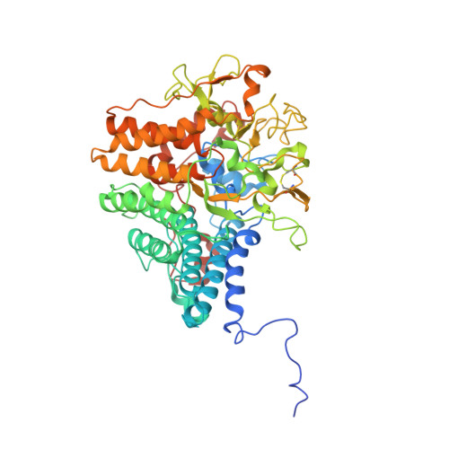
SusD homologA [auth B],
C [auth J]
580Bacteroides thetaiotaomicron VPI-5482Mutation(s): 0 
Gene Names: BT_1762
Membrane Entity: Yes 
UniProt
Find proteins for Q8A6W4 (Bacteroides thetaiotaomicron (strain ATCC 29148 / DSM 2079 / JCM 5827 / CCUG 10774 / NCTC 10582 / VPI-5482 / E50))
Explore Q8A6W4 
Go to UniProtKB:  Q8A6W4
Entity Groups  
Sequence Clusters30% Identity50% Identity70% Identity90% Identity95% Identity100% Identity
UniProt GroupQ8A6W4
Sequence Annotations
Expand
  • Reference Sequence
Find similar proteins by:  (by identity cutoff)  |  3D Structure
Entity ID: 2
MoleculeChains Sequence LengthOrganismDetailsImage
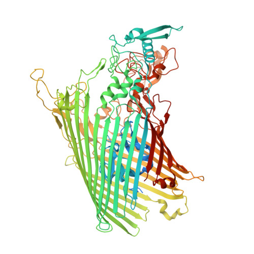
SusC homologB [auth A],
D [auth I]
1,041Bacteroides thetaiotaomicron VPI-5482Mutation(s): 0 
Gene Names: BT_1763
Membrane Entity: Yes 
UniProt
Find proteins for Q8A6W3 (Bacteroides thetaiotaomicron (strain ATCC 29148 / DSM 2079 / JCM 5827 / CCUG 10774 / NCTC 10582 / VPI-5482 / E50))
Explore Q8A6W3 
Go to UniProtKB:  Q8A6W3
Entity Groups  
Sequence Clusters30% Identity50% Identity70% Identity90% Identity95% Identity100% Identity
UniProt GroupQ8A6W3
Sequence Annotations
Expand
  • Reference Sequence
Oligosaccharides

Help

Entity ID: 3
MoleculeChains Length2D Diagram Glycosylation3D Interactions
beta-D-fructofuranose-(2-6)-beta-D-fructofuranose-(2-6)-beta-D-fructofuranose-(2-6)-beta-D-fructofuranoseE [auth D],
F [auth L]
4N/A
Glycosylation Resources
GlyTouCan:  G94951WG
GlyCosmos:  G94951WG
Entity ID: 4
MoleculeChains Length2D Diagram Glycosylation3D Interactions
beta-D-fructofuranose-(2-6)-beta-D-fructofuranose-(2-6)-beta-D-fructofuranose-(2-6)-[beta-D-fructofuranose-(2-1)]beta-D-fructofuranose-(2-6)-beta-D-fructofuranose-(2-6)-beta-D-fructofuranoseG [auth C],
H [auth K]
7N/A
Glycosylation Resources
GlyTouCan:  G35823CA
GlyCosmos:  G35823CA
Experimental Data & Validation

Experimental Data

  • Method: ELECTRON MICROSCOPY
  • Resolution: 2.90 Å
  • Aggregation State: PARTICLE 
  • Reconstruction Method: SINGLE PARTICLE 
EM Software:
TaskSoftware PackageVersion
RECONSTRUCTIONRELION3.1

Structure Validation

View Full Validation Report


Entry History & Funding Information

Deposition Data


Funding OrganizationLocationGrant Number
Wellcome TrustUnited Kingdom215064/Z/18/Z
Wellcome TrustUnited Kingdom214222/Z/18/Z
Wellcome TrustUnited Kingdom108466/Z/15/Z

Revision History  (Full details and data files)

  • Version 1.0: 2023-06-07
    Type: Initial release
  • Version 1.1: 2023-06-21
    Changes: Database references
  • Version 1.2: 2024-11-06
    Changes: Data collection, Structure summary