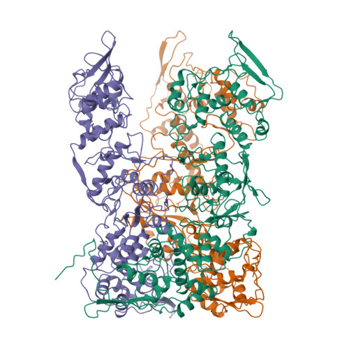
Cryo-ET reveals A10 protein as a major component of the poxvirus palisade layer
Liu, Y., Qu, X., Duan, M., Shi, X., Liu, S., Shi, Y., Gao, G.F.To be published.
Experimental Data Snapshot
Starting Model: in silico
View more details
wwPDB Validation 3D Report Full Report

Entity ID: 1 | |||||
|---|---|---|---|---|---|
| Molecule | Chains | Sequence Length | Organism | Details | Image |
| Core protein OPG136 | 614 | Vaccinia virus WR | Mutation(s): 0 | 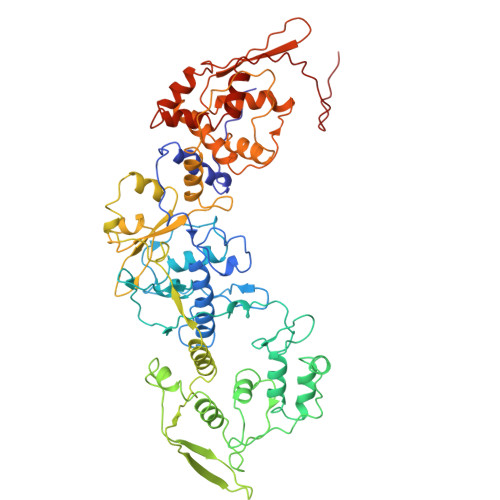 | |
UniProt | |||||
Find proteins for P16715 (Vaccinia virus (strain Western Reserve)) Explore P16715 Go to UniProtKB: P16715 | |||||
Entity Groups | |||||
| Sequence Clusters | 30% Identity50% Identity70% Identity90% Identity95% Identity100% Identity | ||||
| UniProt Group | P16715 | ||||
Sequence AnnotationsExpand | |||||
| |||||
| Task | Software Package | Version |
|---|---|---|
| RECONSTRUCTION | RELION | 3.13 |
RCSB PDB is hosted by

RCSB PDB is a member of the