8SQ7

X-ray crystal structure of Acinetobacter baumanii beta-lactamase variant OXA-82 K83D in complex with doripenem


Experimental Data Snapshot

  • Method: X-RAY DIFFRACTION
  • Resolution: 1.78 Å
  • R-Value Free: 0.181 
  • R-Value Work: 0.155 
  • R-Value Observed: 0.156 

Starting Model: experimental
View more details

wwPDB Validation   3D Report Full Report


Ligand Structure Quality Assessment 


This is version 1.1 of the entry. See complete history


Literature

Structural and Dynamic Features of Acinetobacter baumannii OXA-66 beta-Lactamase Explain Its Stability and Evolution of Novel Variants.

Klamer, Z.L.June, C.M.Wawrzak, Z.Taracila, M.A.Grey, J.A.Benn, A.M.I.Russell, C.P.Bonomo, R.A.Powers, R.A.Leonard, D.A.Szarecka, A.

(2024) J Mol Biol 436: 168603-168603

  • DOI: https://doi.org/10.1016/j.jmb.2024.168603
  • Primary Citation of Related Structures:  
    8SQ7, 8SQ8

  • PubMed Abstract: 

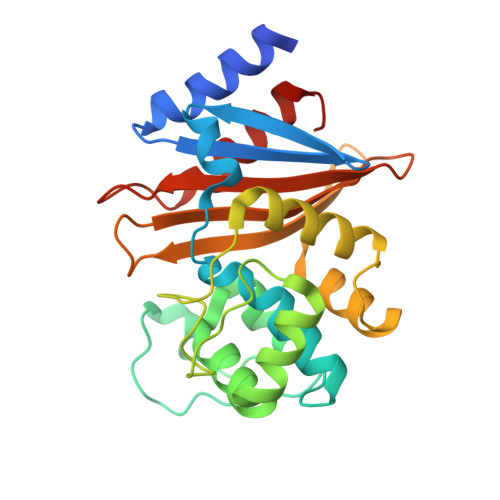
    OXA-66 is a member of the OXA-51 subfamily of class D β-lactamases native to the Acinetobacter genus that includes Acinetobacter baumannii, one of the ESKAPE pathogens and a major cause of drug-resistant nosocomial infections. Although both wild type OXA-66 and OXA-51 have low catalytic activity, they are ubiquitous in the Acinetobacter genomes. OXA-51 is also remarkably thermostable. In addition, newly emerging, single and double amino acid variants show increased activity against carbapenems, indicating that the OXA-51 subfamily is growing and gaining clinical significance. In this study, we used molecular dynamics simulations, X-ray crystallography, and thermal denaturation data to examine and compare the dynamics of OXA-66 wt and its gain-of-function variants: I129L (OXA-83), L167V (OXA-82), P130Q (OXA-109), P130A, and W222L (OXA-234). Our data indicate that OXA-66 wt also has a high melting temperature, and its remarkable stability is due to an extensive and rigid hydrophobic bridge formed by a number of residues around the active site and harbored by the three loops, P, Ω, and β5-β6. Compared to the WT enzyme, the mutants exhibit higher flexibility only in the loop regions, and are more stable than other robust carbapenemases, such as OXA-23 and OXA-24/40. All the mutants show increased rotational flexibility of residues I129 and W222, which allows carbapenems to bind. Overall, our data support the hypothesis that structural features in OXA-51 and OXA-66 promote evolution of multiple highly stable variants with increased clinical relevance in A. baumannii.


  • Organizational Affiliation

    Department of Cell and Molecular Biology, Grand Valley State University, Allendale, MI, USA.


Macromolecules
Find similar proteins by:  (by identity cutoff)  |  3D Structure
Entity ID: 1
MoleculeChains Sequence LengthOrganismDetailsImage
Beta-lactamase OXA-82
A, B, C, D, E
A, B, C, D, E, F, G, H
250Acinetobacter baumanniiMutation(s): 1 
Gene Names: bla-OXA-82blaOXA-82oxa66
UniProt
Find proteins for A8HDA9 (Acinetobacter baumannii)
Explore A8HDA9 
Go to UniProtKB:  A8HDA9
Entity Groups  
Sequence Clusters30% Identity50% Identity70% Identity90% Identity95% Identity100% Identity
UniProt GroupA8HDA9
Sequence Annotations
Expand
  • Reference Sequence
Small Molecules
Ligands 3 Unique
IDChains Name / Formula / InChI Key2D Diagram3D Interactions
4J6 (Subject of Investigation/LOI)
Query on 4J6

Download Ideal Coordinates CCD File 
I [auth A]
K [auth B]
O [auth C]
Q [auth D]
S [auth E]
I [auth A],
K [auth B],
O [auth C],
Q [auth D],
S [auth E],
U [auth F],
W [auth G],
Y [auth H]
(4R,5S)-5-[(2S,3R)-3-hydroxy-1-oxobutan-2-yl]-4-methyl-3-({(3S,5S)-5-[(sulfamoylamino)methyl]pyrrolidin-3-yl}sulfanyl)-4,5-dihydro-1H-pyrrole-2-carboxylic acid
C15 H26 N4 O6 S2
QJKQOMNNBOTMCR-PQTSNVLCSA-N
FLC
Query on FLC

Download Ideal Coordinates CCD File 
J [auth A],
P [auth C],
R [auth D],
T [auth E]
CITRATE ANION
C6 H5 O7
KRKNYBCHXYNGOX-UHFFFAOYSA-K
GOL
Query on GOL

Download Ideal Coordinates CCD File 
AA [auth H]
L [auth B]
M [auth B]
N [auth B]
V [auth F]
AA [auth H],
L [auth B],
M [auth B],
N [auth B],
V [auth F],
X [auth G],
Z [auth H]
GLYCEROL
C3 H8 O3
PEDCQBHIVMGVHV-UHFFFAOYSA-N
Experimental Data & Validation

Experimental Data

  • Method: X-RAY DIFFRACTION
  • Resolution: 1.78 Å
  • R-Value Free: 0.181 
  • R-Value Work: 0.155 
  • R-Value Observed: 0.156 
  • Space Group: P 31
Unit Cell:
Length ( Å )Angle ( ˚ )
a = 70.284α = 90
b = 70.284β = 90
c = 448.355γ = 120
Software Package:
Software NamePurpose
REFMACrefinement
HKL-2000data scaling
HKL-2000data reduction
PHASERphasing

Structure Validation

View Full Validation Report


Ligand Structure Quality Assessment 


Entry History & Funding Information

Deposition Data


Funding OrganizationLocationGrant Number
National Institutes of Health/National Institute Of Allergy and Infectious Diseases (NIH/NIAID)United StatesR01AI072219

Revision History  (Full details and data files)

  • Version 1.0: 2024-05-22
    Type: Initial release
  • Version 1.1: 2024-05-29
    Changes: Database references