8RTT

Structure of the formin Cdc12 bound to the barbed end of phalloidin-stabilized F-actin.


Experimental Data Snapshot

  • Method: ELECTRON MICROSCOPY
  • Resolution: 3.56 Å
  • Aggregation State: PARTICLE 
  • Reconstruction Method: SINGLE PARTICLE 

wwPDB Validation   3D Report Full Report


This is version 1.1 of the entry. See complete history


Literature

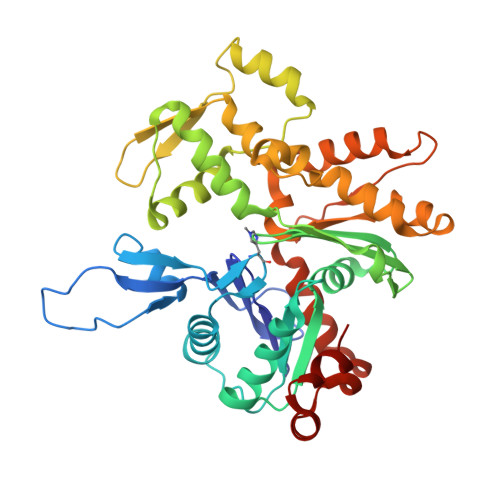
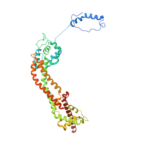
Molecular mechanism of actin filament elongation by formins.

Oosterheert, W.Boiero Sanders, M.Funk, J.Prumbaum, D.Raunser, S.Bieling, P.

(2024) Science 384: eadn9560-eadn9560

  • DOI: https://doi.org/10.1126/science.adn9560
  • Primary Citation of Related Structures:  
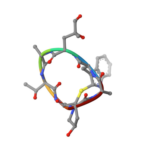
    8RTT, 8RTY, 8RU0, 8RU2, 8RV2

  • PubMed Abstract: 

    Formins control the assembly of actin filaments (F-actin) that drive cell morphogenesis and motility in eukaryotes. However, their molecular interaction with F-actin and their mechanism of action remain unclear. In this work, we present high-resolution cryo-electron microscopy structures of F-actin barbed ends bound by three distinct formins, revealing a common asymmetric formin conformation imposed by the filament. Formation of new intersubunit contacts during actin polymerization sterically displaces formin and triggers its translocation. This "undock-and-lock" mechanism explains how actin-filament growth is coordinated with formin movement. Filament elongation speeds are controlled by the positioning and stability of actin-formin interfaces, which distinguish fast and slow formins. Furthermore, we provide a structure of the actin-formin-profilin ring complex, which resolves how profilin is rapidly released from the barbed end during filament elongation.


  • Organizational Affiliation

    Department of Structural Biochemistry, Max Planck Institute of Molecular Physiology, 44227 Dortmund, Germany.


Macromolecules
Find similar proteins by:  (by identity cutoff)  |  3D Structure
Entity ID: 1
MoleculeChains Sequence LengthOrganismDetailsImage
Actin, cytoplasmic 1, N-terminally processed
A, B, C, D
375Homo sapiensMutation(s): 1 
Gene Names: ACTB
UniProt & NIH Common Fund Data Resources
Find proteins for P60709 (Homo sapiens)
Explore P60709 
Go to UniProtKB:  P60709
PHAROS:  P60709
GTEx:  ENSG00000075624 
Entity Groups  
Sequence Clusters30% Identity50% Identity70% Identity90% Identity95% Identity100% Identity
UniProt GroupP60709
Sequence Annotations
Expand
  • Reference Sequence
Find similar proteins by:  (by identity cutoff)  |  3D Structure
Entity ID: 2
MoleculeChains Sequence LengthOrganismDetailsImage
Cell division control protein 12E,
I [auth F]
419Schizosaccharomyces pombeMutation(s): 0 
Gene Names: cdc12SPAC1F5.04c
UniProt
Find proteins for Q10059 (Schizosaccharomyces pombe (strain 972 / ATCC 24843))
Explore Q10059 
Go to UniProtKB:  Q10059
Entity Groups  
Sequence Clusters30% Identity50% Identity70% Identity90% Identity95% Identity100% Identity
UniProt GroupQ10059
Sequence Annotations
Expand
  • Reference Sequence

Find similar proteins by:  Sequence   |   3D Structure  

Entity ID: 3
MoleculeChains Sequence LengthOrganismDetailsImage
PhalloidinF [auth H],
G [auth I],
H [auth J]
7Amanita phalloidesMutation(s): 0 
Sequence Annotations
Expand
  • Reference Sequence
Small Molecules
Ligands 3 Unique
IDChains Name / Formula / InChI Key2D Diagram3D Interactions
ADP (Subject of Investigation/LOI)
Query on ADP

Download Ideal Coordinates CCD File 
J [auth A],
M [auth B],
P [auth C],
S [auth D]
ADENOSINE-5'-DIPHOSPHATE
C10 H15 N5 O10 P2
XTWYTFMLZFPYCI-KQYNXXCUSA-N
PO4
Query on PO4

Download Ideal Coordinates CCD File 
L [auth A],
O [auth B],
R [auth C]
PHOSPHATE ION
O4 P
NBIIXXVUZAFLBC-UHFFFAOYSA-K
MG
Query on MG

Download Ideal Coordinates CCD File 
K [auth A],
N [auth B],
Q [auth C],
T [auth D]
MAGNESIUM ION
Mg
JLVVSXFLKOJNIY-UHFFFAOYSA-N
Modified Residues  2 Unique
IDChains TypeFormula2D DiagramParent
HIC
Query on HIC
A, B, C, D
L-PEPTIDE LINKINGC7 H11 N3 O2HIS
HYP
Query on HYP
F [auth H],
G [auth I],
H [auth J]
L-PEPTIDE LINKINGC5 H9 N O3PRO
Biologically Interesting Molecules (External Reference) 1 Unique
Experimental Data & Validation

Experimental Data

  • Method: ELECTRON MICROSCOPY
  • Resolution: 3.56 Å
  • Aggregation State: PARTICLE 
  • Reconstruction Method: SINGLE PARTICLE 
EM Software:
TaskSoftware PackageVersion
RECONSTRUCTIONRELION3.1.0
MODEL REFINEMENTCoot0.9.8.1
MODEL REFINEMENTPHENIX1.21rc1_5015

Structure Validation

View Full Validation Report


Entry History & Funding Information

Deposition Data


Funding OrganizationLocationGrant Number
Alexander von Humboldt FoundationGermany--
German Research Foundation (DFG)GermanyBI 1998/2-1
European Research Council (ERC)European Union856118

Revision History  (Full details and data files)

  • Version 1.0: 2024-04-10
    Type: Initial release
  • Version 1.1: 2024-06-05
    Changes: Database references