8ONE

Crystal Structure of full-length Human Lysyl Hydroxylase LH3 - Asp190Ser mutant - Cocrystal with Fe2+, Mn2+, UDP-Glucose


Experimental Data Snapshot

  • Method: X-RAY DIFFRACTION
  • Resolution: 2.30 Å
  • R-Value Free: 0.228 
  • R-Value Work: 0.204 
  • R-Value Observed: 0.205 

Starting Model: experimental
View more details

wwPDB Validation   3D Report Full Report


Ligand Structure Quality Assessment 


This is version 1.2 of the entry. See complete history


Literature

Identification of Regulatory Molecular "Hot Spots" for LH/PLOD Collagen Glycosyltransferase Activity.

Mattoteia, D.Chiapparino, A.Fumagalli, M.De Marco, M.De Giorgi, F.Negro, L.Pinnola, A.Faravelli, S.Roscioli, T.Scietti, L.Forneris, F.

(2023) Int J Mol Sci 24

  • DOI: https://doi.org/10.3390/ijms241311213
  • Primary Citation of Related Structures:  
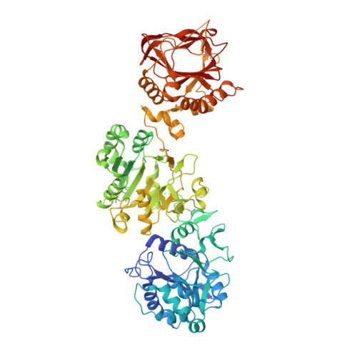
    8ONE

  • PubMed Abstract: 

    Hydroxylysine glycosylations are post-translational modifications (PTMs) essential for the maturation and homeostasis of fibrillar and non-fibrillar collagen molecules. The multifunctional collagen lysyl hydroxylase 3 (LH3/PLOD3) and the collagen galactosyltransferase GLT25D1 are the human enzymes that have been identified as being responsible for the glycosylation of collagen lysines, although a precise description of the contribution of each enzyme to these essential PTMs has not yet been provided in the literature. LH3/PLOD3 is thought to be capable of performing two chemically distinct collagen glycosyltransferase reactions using the same catalytic site: an inverting beta-1,O-galactosylation of hydroxylysines (Gal-T) and a retaining alpha-1,2-glucosylation of galactosyl hydroxylysines (Glc-T). In this work, we have combined indirect luminescence-based assays with direct mass spectrometry-based assays and molecular structure studies to demonstrate that LH3/PLOD3 only has Glc-T activity and that GLT25D1 only has Gal-T activity. Structure-guided mutagenesis confirmed that the Glc-T activity is defined by key residues in the first-shell environment of the glycosyltransferase catalytic site as well as by long-range contributions from residues within the same glycosyltransferase (GT) domain. By solving the molecular structures and characterizing the interactions and solving the molecular structures of human LH3/PLOD3 in complex with different UDP-sugar analogs, we show how these studies could provide insights for LH3/PLOD3 glycosyltransferase inhibitor development. Collectively, our data provide new tools for the direct investigation of collagen hydroxylysine PTMs and a comprehensive overview of the complex network of shapes, charges, and interactions that enable LH3/PLOD3 glycosyltransferase activities, expanding the molecular framework and facilitating an improved understanding and manipulation of glycosyltransferase functions in biomedical applications.


  • Organizational Affiliation

    The Armenise-Harvard Laboratory of Structural Biology, Department of Biology and Biotechnology, University of Pavia, Via Ferrata 9A, 27100 Pavia, Italy.


Macromolecules
Find similar proteins by:  (by identity cutoff)  |  3D Structure
Entity ID: 1
MoleculeChains Sequence LengthOrganismDetailsImage
Multifunctional procollagen lysine hydroxylase and glycosyltransferase LH3719Homo sapiensMutation(s): 1 
Gene Names: PLOD3
EC: 1.14.11.4 (PDB Primary Data), 2.4.1.50 (PDB Primary Data), 2.4.1.66 (PDB Primary Data)
UniProt & NIH Common Fund Data Resources
Find proteins for O60568 (Homo sapiens)
Explore O60568 
Go to UniProtKB:  O60568
PHAROS:  O60568
GTEx:  ENSG00000106397 
Entity Groups  
Sequence Clusters30% Identity50% Identity70% Identity90% Identity95% Identity100% Identity
UniProt GroupO60568
Glycosylation
Glycosylation Sites: 1Go to GlyGen: O60568-1
Sequence Annotations
Expand
  • Reference Sequence
Oligosaccharides

Help

Entity ID: 2
MoleculeChains Length2D Diagram Glycosylation3D Interactions
2-acetamido-2-deoxy-beta-D-glucopyranose-(1-4)-2-acetamido-2-deoxy-beta-D-glucopyranoseB [auth L]2N/A
Glycosylation Resources
GlyTouCan:  G42666HT
GlyCosmos:  G42666HT
GlyGen:  G42666HT
Small Molecules
Ligands 6 Unique
IDChains Name / Formula / InChI Key2D Diagram3D Interactions
UPG (Subject of Investigation/LOI)
Query on UPG

Download Ideal Coordinates CCD File 
K [auth A]URIDINE-5'-DIPHOSPHATE-GLUCOSE
C15 H24 N2 O17 P2
HSCJRCZFDFQWRP-JZMIEXBBSA-N
NAG
Query on NAG

Download Ideal Coordinates CCD File 
G [auth A]2-acetamido-2-deoxy-beta-D-glucopyranose
C8 H15 N O6
OVRNDRQMDRJTHS-FMDGEEDCSA-N
AKG
Query on AKG

Download Ideal Coordinates CCD File 
C [auth A]2-OXOGLUTARIC ACID
C5 H6 O5
KPGXRSRHYNQIFN-UHFFFAOYSA-N
GOL
Query on GOL

Download Ideal Coordinates CCD File 
H [auth A],
I [auth A],
J [auth A]
GLYCEROL
C3 H8 O3
PEDCQBHIVMGVHV-UHFFFAOYSA-N
FE2
Query on FE2

Download Ideal Coordinates CCD File 
D [auth A],
E [auth A]
FE (II) ION
Fe
CWYNVVGOOAEACU-UHFFFAOYSA-N
MN
Query on MN

Download Ideal Coordinates CCD File 
F [auth A]MANGANESE (II) ION
Mn
WAEMQWOKJMHJLA-UHFFFAOYSA-N
Experimental Data & Validation

Experimental Data

  • Method: X-RAY DIFFRACTION
  • Resolution: 2.30 Å
  • R-Value Free: 0.228 
  • R-Value Work: 0.204 
  • R-Value Observed: 0.205 
  • Space Group: C 2 2 21
Unit Cell:
Length ( Å )Angle ( ˚ )
a = 97.09α = 90
b = 100.181β = 90
c = 223.794γ = 90
Software Package:
Software NamePurpose
PHENIXrefinement
XDSdata reduction
Aimlessdata scaling
PHASERphasing

Structure Validation

View Full Validation Report


Ligand Structure Quality Assessment 


Entry History & Funding Information

Deposition Data


Funding OrganizationLocationGrant Number
The Giovanni Armenise-Harvard FoundationUnited StatesCDA 2013
Italian Association for Cancer ResearchItalyMFAG 20075
Italian Association for Cancer ResearchItalyBridge 27004
Mizutani Foundation for GlycoscienceJapan200039

Revision History  (Full details and data files)

  • Version 1.0: 2023-07-19
    Type: Initial release
  • Version 1.1: 2023-07-26
    Changes: Database references
  • Version 1.2: 2023-08-09
    Changes: Derived calculations