8ISP

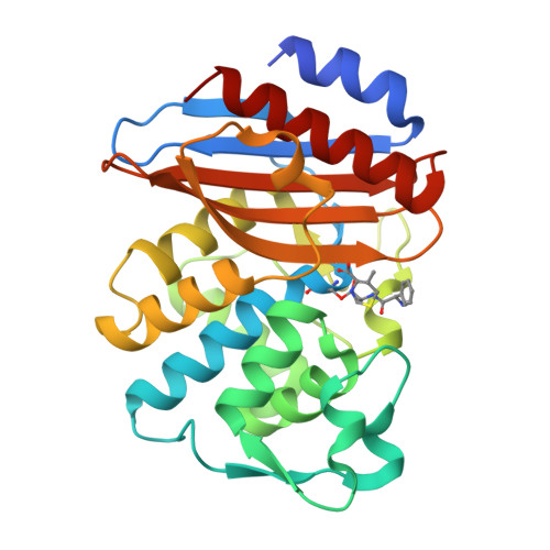
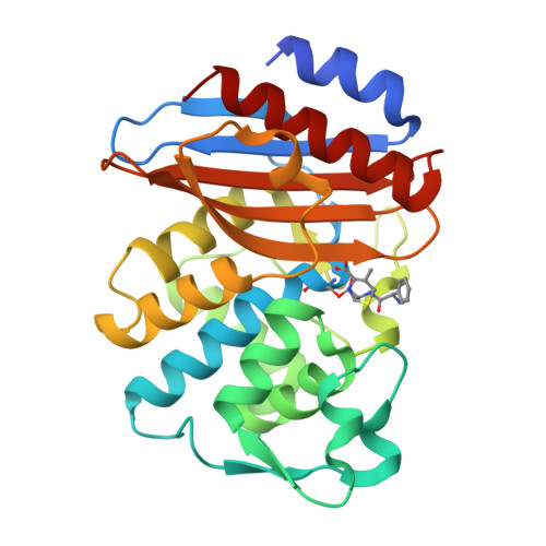
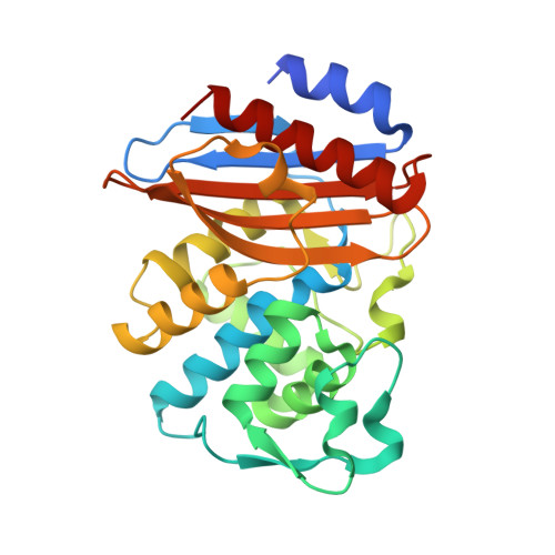
Crystal structure of extended-spectrum class A beta-lactamase, CESS-1 E166Q acylated by cephalexin


Experimental Data Snapshot

  • Method: X-RAY DIFFRACTION
  • Resolution: 2.11 Å
  • R-Value Free: 0.243 
  • R-Value Work: 0.193 
  • R-Value Observed: 0.198 

Starting Model: experimental
View more details

wwPDB Validation   3D Report Full Report


Ligand Structure Quality Assessment 

Created with Raphaël 2.3.0Worse 01 BetterLigand structure goodness of fit to experimental dataBest fitted Q53Click on this verticalbar to view details

This is version 1.1 of the entry. See complete history


Literature

Characterization of the extended substrate spectrum of the class A beta-lactamase CESS-1 from Stenotrophomonas sp. and structure-based investigation into its substrate preference.

Jeong, B.G.Kim, M.Y.Jeong, C.S.Do, H.Hwang, J.Lee, J.H.Cha, S.S.

(2024) Int J Antimicrob Agents 63: 107171-107171

  • DOI: https://doi.org/10.1016/j.ijantimicag.2024.107171
  • Primary Citation of Related Structures:  
    8ISO, 8ISP, 8ISQ, 8ISR

  • PubMed Abstract: 

    Stenotrophomonas spp. intrinsically resistant to many β-lactam antibiotics are found throughout the environment. CESS-1 identified in Stenotrophomonas sp. KCTC 12332 is an uncharacterized class A β-lactamase. The goal of this study was to reveal biochemical and structural characteristics of CESS-1. The hydrolytic activities of CESS-1 towards penicillins (penicillin G and ampicillin), cephalosporins (cephalexin, cefaclor, and cefotaxime), and carbapenems (imipenem and meropenem) was spectrophotometrically monitored. Structural information on E166Q mutants of CESS-1 acylated by cefaclor, cephalexin, or ampicillin were determined by X-ray crystallography. CESS-1 displayed hydrolytic activities toward penicillins and cephalosporins, with negligible activity toward carbapenems. Although cefaclor, cephalexin, and ampicillin have similar structures with identical R1 side chains, the catalytic parameters of CESS-1 toward them were distinct. The k cat values for cefaclor, cephalexin, and ampicillin were 1249.6 s -1 , 204.3 s -1 , and 69.8 s -1 , respectively, with the accompanying K M values of 287.6 μM, 236.7 μM, and 28.8 μM, respectively. CESS-1 was able to discriminate between cefaclor and cephalexin with a single structural difference at C3 position: -Cl (cefaclor) and -CH 3 (cephalexin). Structural comparisons among three E166Q mutants of CESS-1 acylated by cefaclor, cephalexin, or ampicillin, revealed that cooperative positional changes in the R1 side chain of substrates and their interaction with the β5-β6 loop affect the distance between Asn170 and the deacylating water at the acyl-enzyme intermediate state. This is directly associated with the differential hydrolytic activities of CESS-1 toward the three structurally similar β-lactam antibiotics.


  • Organizational Affiliation

    Department of Chemistry & Nanoscience, Ewha Womans University, Seoul, Republic of Korea.


Macromolecules
Find similar proteins by:  (by identity cutoff)  |  3D Structure
Entity ID: 1
MoleculeChains Sequence LengthOrganismDetailsImage
Beta-lactamase269Stenotrophomonas sp. KCTC 12332Mutation(s): 1 
Gene Names: AXG53_04720
EC: 3.5.2.6
UniProt
Find proteins for A0A126NGE0 (Stenotrophomonas sp. KCTC 12332)
Explore A0A126NGE0 
Go to UniProtKB:  A0A126NGE0
Entity Groups  
Sequence Clusters30% Identity50% Identity70% Identity90% Identity95% Identity100% Identity
UniProt GroupA0A126NGE0
Sequence Annotations
Expand
  • Reference Sequence
Small Molecules
Ligands 1 Unique
IDChains Name / Formula / InChI Key2D Diagram3D Interactions
Q53 (Subject of Investigation/LOI)
Query on Q53

Download Ideal Coordinates CCD File 
B [auth A] (R)-2-((R)-((R)-2-amino-2-phenylacetamido)(carboxy)methyl)-5-methyl-3,6-dihydro-2H-1,3-thiazine-4-carboxylic acid
C16 H19 N3 O5 S
MSGWVKZSYIMFHD-SCDSUCTJSA-N
Experimental Data & Validation

Experimental Data

  • Method: X-RAY DIFFRACTION
  • Resolution: 2.11 Å
  • R-Value Free: 0.243 
  • R-Value Work: 0.193 
  • R-Value Observed: 0.198 
  • Space Group: P 41 21 2
Unit Cell:
Length ( Å )Angle ( ˚ )
a = 62.118α = 90
b = 62.118β = 90
c = 150.448γ = 90
Software Package:
Software NamePurpose
PHENIXrefinement
HKL-2000data collection
Cootmodel building
PHASERphasing
HKL-2000data reduction

Structure Validation

View Full Validation Report


Ligand Structure Quality Assessment 

Created with Raphaël 2.3.0Worse 01 BetterLigand structure goodness of fit to experimental dataBest fitted Q53Click on this verticalbar to view details

Entry History & Funding Information

Deposition Data


Funding OrganizationLocationGrant Number
Not funded--

Revision History  (Full details and data files)

  • Version 1.0: 2024-05-15
    Type: Initial release
  • Version 1.1: 2024-10-16
    Changes: Structure summary