8IN9

The structure of the GfsA KSQ-AT didomain in complex with the GfsA ACP domain


Experimental Data Snapshot

  • Method: X-RAY DIFFRACTION
  • Resolution: 3.40 Å
  • R-Value Free: 
    0.311 (Depositor), 0.310 (DCC) 
  • R-Value Work: 
    0.265 (Depositor), 0.270 (DCC) 
  • R-Value Observed: 
    0.267 (Depositor) 

Starting Models: in silico, experimental
View more details

wwPDB Validation   3D Report Full Report


Ligand Structure Quality Assessment 

Created with Raphaël 2.3.0Worse 01 BetterLigand structure goodness of fit to experimental dataBest fitted 9EFClick on this verticalbar to view details

This is version 1.2 of the entry. See complete history


Literature

Structure-Based Analysis of Transient Interactions between Ketosynthase-like Decarboxylase and Acyl Carrier Protein in a Loading Module of Modular Polyketide Synthase.

Chisuga, T.Murakami, S.Miyanaga, A.Kudo, F.Eguchi, T.

(2023) ACS Chem Biol 18: 1398-1404

  • DOI: https://doi.org/10.1021/acschembio.3c00151
  • Primary Citation of Related Structures:  
    8IN9

  • PubMed Abstract: 

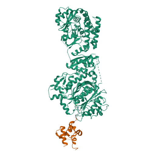
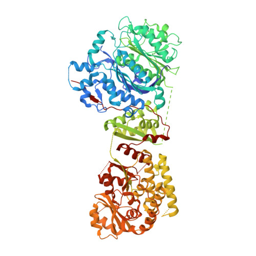
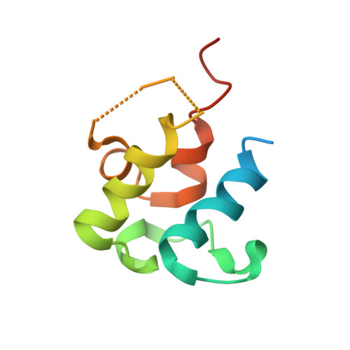
    Ketosynthase-like decarboxylase (KS Q ) domains are widely distributed in the loading modules of modular type I polyketide synthases (PKSs) and catalyze the decarboxylation of the (alkyl-)malonyl unit bound to the acyl carrier protein (ACP) in the loading module for the construction of the PKS starter unit. Previously, we performed a structural and functional analysis of the GfsA KS Q domain involved in the biosynthesis of macrolide antibiotic FD-891. We furthermore revealed the recognition mechanism for the malonic acid thioester moiety of the malonyl-GfsA loading module ACP (ACP L ) as a substrate. However, the exact recognition mechanism for the GfsA ACP L moiety remains unclear. Here, we present a structural basis for the interactions between the GfsA KS Q domain and GfsA ACP L . We determined the crystal structure of the GfsA KS Q -acyltransferase (AT) didomain in complex with ACP L (ACP L =KS Q AT complex) by using a pantetheine crosslinking probe. We identified the key amino acid residues involved in the KS Q domain-ACP L interactions and confirmed the importance of these residues by mutational analysis. The binding mode of ACP L to the GfsA KS Q domain is similar to that of ACP to the ketosynthase domain in modular type I PKSs. Furthermore, comparing the ACP L =KS Q AT complex structure with other full-length PKS module structures provides important insights into the overall architectures and conformational dynamics of the type I PKS modules.


  • Organizational Affiliation

    Department of Chemistry, Tokyo Institute of Technology, 2-12-1 O-Okayama, Tokyo 152-8551, Japan.


Macromolecules
Find similar proteins by:  (by identity cutoff)  |  3D Structure
Entity ID: 1
MoleculeChains Sequence LengthOrganismDetailsImage
Polyketide synthase916Streptomyces graminofaciensMutation(s): 0 
Gene Names: gfsA
EC: 2.3.1 (UniProt), 4.1.1 (UniProt)
UniProt
Find proteins for E0D202 (Streptomyces halstedii)
Explore E0D202 
Go to UniProtKB:  E0D202
Entity Groups  
Sequence Clusters30% Identity50% Identity70% Identity90% Identity95% Identity100% Identity
UniProt GroupE0D202
Sequence Annotations
Expand
  • Reference Sequence
Find similar proteins by:  (by identity cutoff)  |  3D Structure
Entity ID: 2
MoleculeChains Sequence LengthOrganismDetailsImage
Polyketide synthase94Streptomyces graminofaciensMutation(s): 0 
Gene Names: gfsA
EC: 2.3.1 (UniProt), 4.1.1 (UniProt)
UniProt
Find proteins for E0D202 (Streptomyces halstedii)
Explore E0D202 
Go to UniProtKB:  E0D202
Entity Groups  
Sequence Clusters30% Identity50% Identity70% Identity90% Identity95% Identity100% Identity
UniProt GroupE0D202
Sequence Annotations
Expand
  • Reference Sequence
Small Molecules
Ligands 1 Unique
IDChains Name / Formula / InChI Key2D Diagram3D Interactions
9EF (Subject of Investigation/LOI)
Query on 9EF

Download Ideal Coordinates CCD File 
C [auth A]N-[2-(acetylamino)ethyl]-N~3~-[(2R)-2-hydroxy-3,3-dimethyl-4-(phosphonooxy)butanoyl]-beta-alaninamide
C13 H26 N3 O8 P
GTANJSAWPNEGPZ-NSHDSACASA-N
Experimental Data & Validation

Experimental Data

  • Method: X-RAY DIFFRACTION
  • Resolution: 3.40 Å
  • R-Value Free:  0.311 (Depositor), 0.310 (DCC) 
  • R-Value Work:  0.265 (Depositor), 0.270 (DCC) 
  • R-Value Observed: 0.267 (Depositor) 
Space Group: I 41 2 2
Unit Cell:
Length ( Å )Angle ( ˚ )
a = 102.212α = 90
b = 102.212β = 90
c = 408.013γ = 90
Software Package:
Software NamePurpose
PHENIXrefinement
XDSdata reduction
Aimlessdata scaling
MOLREPphasing
Cootmodel building

Structure Validation

View Full Validation Report


Ligand Structure Quality Assessment 

Created with Raphaël 2.3.0Worse 01 BetterLigand structure goodness of fit to experimental dataBest fitted 9EFClick on this verticalbar to view details

Entry History & Funding Information

Deposition Data


Funding OrganizationLocationGrant Number
Japan Society for the Promotion of Science (JSPS)Japan20H02911

Revision History  (Full details and data files)

  • Version 1.0: 2023-05-31
    Type: Initial release
  • Version 1.1: 2023-06-28
    Changes: Database references
  • Version 1.2: 2024-11-20
    Changes: Data collection, Structure summary