8ILH

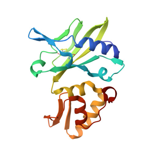
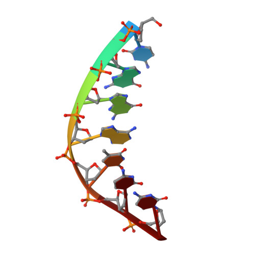
The crystal structure of dG(Se-Sp)-DNA:Pol X product binary complex


Experimental Data Snapshot

  • Method: X-RAY DIFFRACTION
  • Resolution: 2.10 Å
  • R-Value Free: 0.227 
  • R-Value Work: 0.181 
  • R-Value Observed: 0.183 

Starting Model: experimental
View more details

wwPDB Validation   3D Report Full Report


Ligand Structure Quality Assessment 


This is version 2.0 of the entry. See complete history


Literature

Structural Insight into Polymerase Mechanism via a Chiral Center Generated with a Single Selenium Atom.

Qin, T.Hu, B.Zhao, Q.Wang, Y.Wang, S.Luo, D.Lyu, J.Chen, Y.Gan, J.Huang, Z.

(2023) Int J Mol Sci 24

  • DOI: https://doi.org/10.3390/ijms242115758
  • Primary Citation of Related Structures:  
    8ILD, 8ILE, 8ILF, 8ILG, 8ILH, 8ILI

  • PubMed Abstract: 

    DNA synthesis catalyzed by DNA polymerase is essential for all life forms, and phosphodiester bond formation with phosphorus center inversion is a key step in this process. Herein, by using a single-selenium-atom-modified dNTP probe, we report a novel strategy to visualize the reaction stereochemistry and catalysis. We capture the before- and after-reaction states and provide explicit evidence of the center inversion and in-line attacking S N 2 mechanism of DNA polymerization, while solving the diastereomer absolute configurations. Further, our kinetic and thermodynamic studies demonstrate that in the presence of Mg 2+ ions (or Mn 2+ ), the binding affinity ( K m ) and reaction selectivity ( k cat / K m ) of dGTPαSe-Rp were 51.1-fold (or 19.5-fold) stronger and 21.8-fold (or 11.3-fold) higher than those of dGTPαSe-Sp, respectively, indicating that the diastereomeric Se-Sp atom was quite disruptive of the binding and catalysis. Our findings reveal that the third metal ion is much more critical than the other two metal ions in both substrate recognition and bond formation, providing insights into how to better design the polymerase inhibitors and discover the therapeutics.


  • Organizational Affiliation

    Key Laboratory of Bio-Resource and Eco-Environment of Ministry of Education, College of Life Sciences, Sichuan University, Chengdu 610064, China.


Macromolecules

Find similar proteins by:  (by identity cutoff)  |  3D Structure
Entity ID: 1
MoleculeChains Sequence LengthOrganismDetailsImage
Repair DNA polymerase X
A, B, C, D
177African swine fever virus BA71VMutation(s): 0 
Gene Names: Ba71V-97O174L
EC: 2.7.7.7
UniProt
Find proteins for P42494 (African swine fever virus (strain Badajoz 1971 Vero-adapted))
Explore P42494 
Go to UniProtKB:  P42494
Entity Groups  
Sequence Clusters30% Identity50% Identity70% Identity90% Identity95% Identity100% Identity
UniProt GroupP42494
Sequence Annotations
Expand
  • Reference Sequence

Find similar nucleic acids by:  Sequence   |   3D Structure  

Entity ID: 2
MoleculeChains LengthOrganismImage
DNA (5'-D(*CP*GP*GP*AP*TP*CP*C)-3')
E, F, G, H
7synthetic construct
Sequence Annotations
Expand
  • Reference Sequence
Small Molecules
Ligands 4 Unique
IDChains Name / Formula / InChI Key2D Diagram3D Interactions
8EB (Subject of Investigation/LOI)
Query on 8EB

Download Ideal Coordinates CCD File 
N [auth B][[(2R,3S,5R)-5-(2-azanyl-6-oxidanylidene-3H-purin-9-yl)-3-oxidanyl-oxolan-2-yl]methoxy-selanyl-phosphoryl] dihydrogen phosphate
C10 H15 N5 O9 P2 Se
XCFKPSIHHMJWLO-PDRQRWKTSA-N
GMS (Subject of Investigation/LOI)
Query on GMS

Download Ideal Coordinates CCD File 
AA [auth G],
BA [auth H],
Z [auth E]
2'-DEOXYGUANOSINE-5'-MONOSELENOPHOSPHATE
C10 H14 N5 O6 P Se
MMKHSOCVUGNOOR-KVQBGUIXSA-N
SO4
Query on SO4

Download Ideal Coordinates CCD File 
K [auth A]
L [auth A]
M [auth A]
Q [auth B]
T [auth C]
K [auth A],
L [auth A],
M [auth A],
Q [auth B],
T [auth C],
U [auth C],
X [auth D],
Y [auth D]
SULFATE ION
O4 S
QAOWNCQODCNURD-UHFFFAOYSA-L
MG
Query on MG

Download Ideal Coordinates CCD File 
I [auth A]
J [auth A]
O [auth B]
P [auth B]
R [auth C]
I [auth A],
J [auth A],
O [auth B],
P [auth B],
R [auth C],
S [auth C],
V [auth D],
W [auth D]
MAGNESIUM ION
Mg
JLVVSXFLKOJNIY-UHFFFAOYSA-N
Experimental Data & Validation

Experimental Data

  • Method: X-RAY DIFFRACTION
  • Resolution: 2.10 Å
  • R-Value Free: 0.227 
  • R-Value Work: 0.181 
  • R-Value Observed: 0.183 
  • Space Group: P 1 21 1
Unit Cell:
Length ( Å )Angle ( ˚ )
a = 60.471α = 90
b = 87.28β = 91.15
c = 87.056γ = 90
Software Package:
Software NamePurpose
REFMACrefinement
XDSdata reduction
HKL-2000data scaling
PHENIXphasing

Structure Validation

View Full Validation Report


Ligand Structure Quality Assessment 


Entry History & Funding Information

Deposition Data


Funding OrganizationLocationGrant Number
Ministry of Science and Technology (MoST, China)China2022YFC2303700
National Natural Science Foundation of China (NSFC)China22107079
National Natural Science Foundation of China (NSFC)China22077089
Ministry of Education (MoE, China)China2020M673205

Revision History  (Full details and data files)

  • Version 1.0: 2024-01-10
    Type: Initial release
  • Version 2.0: 2024-01-17
    Changes: Atomic model, Author supporting evidence, Data collection, Derived calculations, Non-polymer description, Structure summary