8EFY

Structure of double homo-hexameric AAA+ ATPase RuvB motors


Experimental Data Snapshot

  • Method: ELECTRON MICROSCOPY
  • Resolution: 3.16 Å
  • Aggregation State: PARTICLE 
  • Reconstruction Method: SINGLE PARTICLE 

wwPDB Validation   3D Report Full Report


This is version 2.0 of the entry. See complete history


Literature

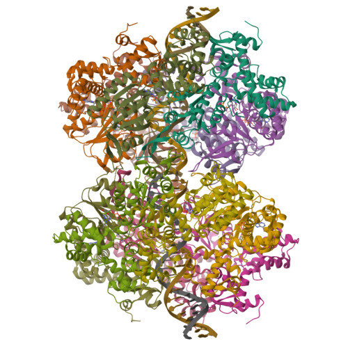
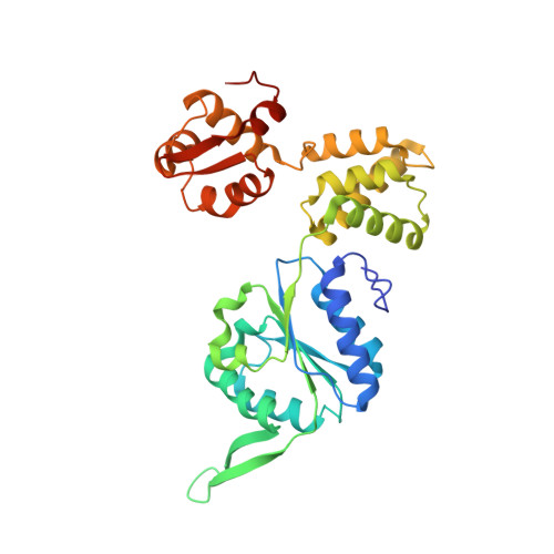
Molecular mechanisms of Holliday junction branch migration catalyzed by an asymmetric RuvB hexamer.

Rish, A.D.Shen, Z.Chen, Z.Zhang, N.Zheng, Q.Fu, T.M.

(2023) Nat Commun 14: 3549-3549

  • DOI: https://doi.org/10.1038/s41467-023-39250-6
  • Primary Citation of Related Structures:  
    8EFV, 8EFY, 8GH8

  • PubMed Abstract: 

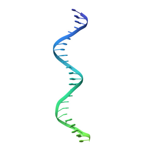
    The Holliday junction (HJ) is a DNA intermediate of homologous recombination, involved in many fundamental physiological processes. RuvB, an ATPase motor protein, drives branch migration of the Holliday junction with a mechanism that had yet to be elucidated. Here we report two cryo-EM structures of RuvB, providing a comprehensive understanding of HJ branch migration. RuvB assembles into a spiral staircase, ring-like hexamer, encircling dsDNA. Four protomers of RuvB contact the DNA backbone with a translocation step size of 2 nucleotides. The variation of nucleotide-binding states in RuvB supports a sequential model for ATP hydrolysis and nucleotide recycling, which occur at separate, singular positions. RuvB's asymmetric assembly also explains the 6:4 stoichiometry between the RuvB/RuvA complex, which coordinates HJ migration in bacteria. Taken together, we provide a mechanistic understanding of HJ branch migration facilitated by RuvB, which may be universally shared by prokaryotic and eukaryotic organisms.


  • Organizational Affiliation

    The Ohio State Biochemistry Program, The Ohio State University, Columbus, OH, 43210, USA.


Macromolecules

Find similar proteins by:  (by identity cutoff)  |  3D Structure
Entity ID: 1
MoleculeChains Sequence LengthOrganismDetailsImage
Holliday junction ATP-dependent DNA helicase RuvB
A, B, C, D, E
324Thermus thermophilus HB8Mutation(s): 0 
Gene Names: ruvBTTHA0406
EC: 3.6.4.12 (PDB Primary Data), 3.6.4 (UniProt)
UniProt
Find proteins for Q5SL87 (Thermus thermophilus (strain ATCC 27634 / DSM 579 / HB8))
Explore Q5SL87 
Go to UniProtKB:  Q5SL87
Entity Groups  
Sequence Clusters30% Identity50% Identity70% Identity90% Identity95% Identity100% Identity
UniProt GroupQ5SL87
Sequence Annotations
Expand
  • Reference Sequence
Small Molecules
Ligands 3 Unique
IDChains Name / Formula / InChI Key2D Diagram3D Interactions
AGS (Subject of Investigation/LOI)
Query on AGS

Download Ideal Coordinates CCD File 
CA [auth J]
EA [auth J]
GA [auth L]
S [auth B]
U [auth B]
PHOSPHOTHIOPHOSPHORIC ACID-ADENYLATE ESTER
C10 H16 N5 O12 P3 S
NLTUCYMLOPLUHL-KQYNXXCUSA-N
ADP (Subject of Investigation/LOI)
Query on ADP

Download Ideal Coordinates CCD File 
AA [auth I]
IA [auth M]
JA [auth N]
Q [auth A]
Y [auth E]
ADENOSINE-5'-DIPHOSPHATE
C10 H15 N5 O10 P2
XTWYTFMLZFPYCI-KQYNXXCUSA-N
MG (Subject of Investigation/LOI)
Query on MG

Download Ideal Coordinates CCD File 
BA [auth I]
DA [auth J]
FA [auth K]
HA [auth L]
R [auth A]
MAGNESIUM ION
Mg
JLVVSXFLKOJNIY-UHFFFAOYSA-N
Experimental Data & Validation

Experimental Data

  • Method: ELECTRON MICROSCOPY
  • Resolution: 3.16 Å
  • Aggregation State: PARTICLE 
  • Reconstruction Method: SINGLE PARTICLE 

Structure Validation

View Full Validation Report


Entry History & Funding Information

Deposition Data


Funding OrganizationLocationGrant Number
Not funded--

Revision History  (Full details and data files)

  • Version 1.0: 2023-05-10
    Type: Initial release
  • Version 2.0: 2024-11-20
    Changes: Advisory, Atomic model, Data collection, Database references, Derived calculations, Structure summary